Pancreatic cyst in a cat: diagnosis and excision
Der Praktische Tierarzt 104, 338-342
DOI: 10.2376/0032-681X-2312
© Schlütersche Fachmedien GmbH. 2023
Eingereicht: 6. September 2022
Akzeptiert: 24. Januar 2022
Publiziert: 04/2023
Zusammenfassung
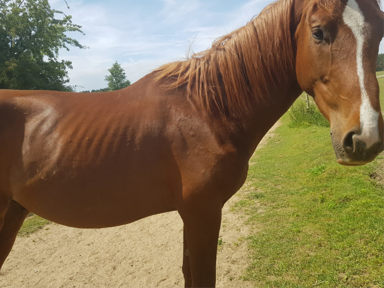
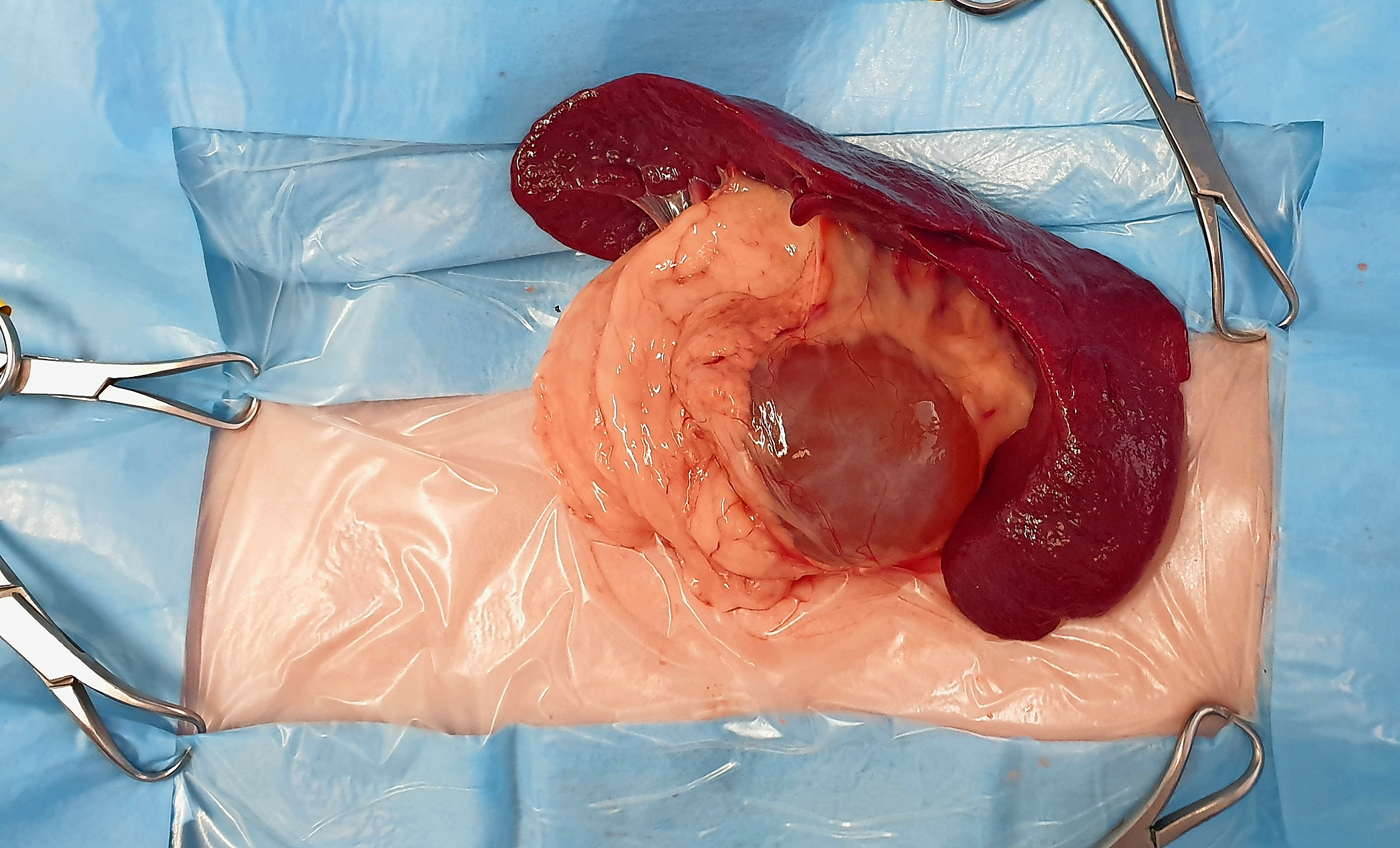
Eine 13 Jahre alte Katze wurde wegen chronischen Gewichtsverlusts in der Klinik vorgestellt. Die klinische Untersuchung war, abgesehen von einem Body Condition Score von 4/9 und einem stumpfen Fell, unauffällig. Die biochemischen Blutuntersuchungen zeigten Hinweise auf eine chronische Pankreatitis. Sonografisch wurde eine zystische Struktur im linken Pankreasschenkel festgestellt. Zur Diagnosestellung und als therapeutische Maßnahme wurden eine chirurgische Exploration der Bauchhöhle und die Resektion der zystischen Struktur mittels partieller, linksseitiger Pankreatektomie durchgeführt. Zudem wurden Bioptate von Magen, Dünndarm und den mesenterialen Lymphknoten entnommen. Alle Proben wurden zur histopathologischen Untersuchung eingesandt. Die histopathologischen Untersuchungen ergaben die Diagnose einer Pankreaszyste und eine geringgradige lymphoplasmazelluläre Enteritis und Gastritis sowie eine reaktive Hyperplasie der Lymphknoten. Die Katze zeigte eine komplikationslose Rehabilitationsphase und im Anschluss ein ungestörtes Allgemeinbefinden mit adäquater Gewichtszunahme über die nächsten Wochen ohne spezifische Therapie. Auf telefonische Nachfrage nach vier und fünf Monaten war der Patient nach wie vor symptomfrei. Es konnten jedoch keine Ultraschallkontrollen durchgeführt werden, um ein mögliches Rezidiv der Zyste sicher auszuschließen.
Summary
A 13-year-old cat was presented due to chronic weight loss. A physical exam was unremarkable besides a body condition score of 4/9 and a dull hair coat. Serum biochemistry results were suspicious for chronic pancreatitis. An abdominal ultrasound examination revealed a cystic lesion in the left lobe of the pancreas. A diagnostic abdominal exploration was performed, including removal of the cyst via a leftsided partial pancreatectomy and biopsies of the stomach, small intestine and the mesenteric lymph nodes were taken. All samples were sent for histopathological evaluation. Histopathological diagnosis was pancreatic cyst and mild lymphoplasmacellular enteritis and gastritis, as well as reactive hyperplasia of the lymph nodes. Recovery was uneventful and over the following weeks the cat was gaining some weight without any specific therapy. The owners reported that the cat was doing well during telephone follow-ups at four and five months post-operatively. Unfortunately, no follow-up ultrasound examinations could be performed to definitely rule out possible recurrence of the cystic lesion.