

Malignes epitheloides Mesotheliom bei einem Prinz-Alfred-Hirsch (Cervus alfredi)
Berliner und Münchener Tierärztliche Wochenschrift 135, 1–6
DOI: 10.2376/1439-0299-2021-28
© Schlütersche Fachmedien GmbH. 2022
Eingereicht: 21. Dezember 2021
Akzeptiert: 21. Januar 2022
Publiziert: 02/2022
Summary
A Visayan spotted deer with progressive weight loss showed diffuse thickening and nodular changes of the pleura and peritoneum. Histology revealed a highly pleomorphic, predominantly solid neoplasm, consistent with a malignant epithelioid mesothelioma. By immunohistochemistry, expression of pan-cytokeratin, cytokeratin 5/6, and vimentin was found in tumor cells. In addition, scattered expression of calretinin, desmin and α-smooth muscle actin was present within the neoplasm. Mesotheliomas should be considered as a potential differential diagnosis for proliferative processes involving the serosa in cervids.
Zusammenfassung
Bei der Sektion eines Prinz-Alfred-Hirschs mit progressivem Gewichtsverlust wurden multiple noduläre, teils konfluierende Herde auf den serösen Häuten der Brust- und Bauchhöhle festgestellt. Histologisch konnte ein malignes, epitheloides Mesotheliom, überwiegend vom soliden Subtyp, nachgewiesen werden. Die Tumorzellen zeigten einen deutlichen Pleomorphismus mit hoher Mitoserate. Mittels Immunhistologie fand sich eine Expression von Pan-Zytokeratin, Zytokeratin 5/6 und Vimentin in den Tumorzellen. Zusätzlich zeigten einzelne neoplastische Zellen eine Expression von Calretinin, Desmin und α-smooth muscle actin. Mesotheliome sollten als Differenzialdiagnose proliferativer Prozesse, welche die Serosa betreffen, bei Cerviden berücksichtigt werden.
Introduction
Mesotheliomas are rare neoplasms in domestic and wildlife animals, originating from the mesothelium of the pericardium, pleura or peritoneum (Plummer 1956, Dukes et al. 1982, Harbison and Godleski 1983). So far, two cases of mesotheliomas in cervids, a sika deer (Cervus nippon yesoensis) and a European spotted fallow deer (Dana dama), have been reported (Byerly et al. 1989, Matsuda et al. 2019). A causative link between asbestos fibers and mesotheliomas is widely accepted in humans (Selikoff et al. 1964). However, tumor development in animals is usually not related to asbestos exposure and no specific etiology can be identified for most mesotheliomas in veterinary medicine. Besides spontaneous occurrences, congenital mesotheliomas have been observed in fetal or young cattle (Misdorp 2002, Munday et al. 2016).
Mesotheliomas cause unspecific signs, such as anorexia, vomiting, and hemorrhages, as well as thoracic and abdominal effusions. They may occur at any serosal lining of body cavities, namely the pericardium, thoracic cavity, abdominal cavity or cavum vaginale. Multicentric mesotheliomas involve multiple locations simultaneously. Macroscopically, mesotheliomas present as diffuse thickening of the serosa or multiple nodules of white-yellowish color and soft to firm consistency (Munday et al. 2016).
Histologically, mesotheliomas can be divided into three categories, namely epithelioid, sarcomatous (spindle-shaped), and biphasic (mixed) tumors. In humans, seventeen different subtypes of epithelioid mesotheliomas have been identified, of which papillary, tubular, solid, sclerosing, cystic, and deciduoid subtypes can be found in animals (Head et al. 2003, Munday et al. 2016). Virtually all mesotheliomas can spread within body cavities by implantation and less commonly by lymphatic metastasis (Stoica et al. 2004, Matsuda et al. 2019).
Top Job:
Adenocarcinomas of the ovary, pancreas, lung, intestine and uterus, as well as the urinary transitional cell carcinoma are important differential diagnoses for mesotheliomas. In addition, non-neoplastic etiologies, such as peritoneal tuberculosis (pearl disease) and other unspecific granulomatous peritonitis need to be differentiated from this entity. To the best of the author`s knowledge, this is the first report of a malignant mesothelioma in a Visayan spotted deer.
Case description
Clinical findings
The 13-year-old, female Visayan spotted deer from a wildlife conservation center showed anorexia and progressive weight loss. The animal received medical treatment (antibiotic and anti-inflammatory medication) over a period of two weeks. Due to unresponsiveness to treatment and worsening of clinical signs, the animal was euthanized and submitted for necropsy at the Department of Pathology of the University of Veterinary Medicine Hanover.
Gross pathology
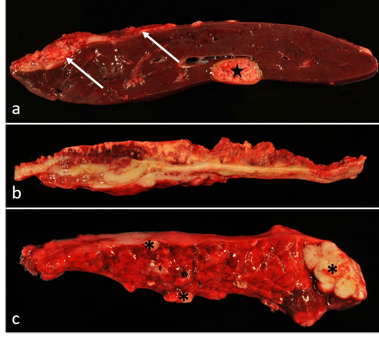
Necropsy was performed and tissues were taken for histologic examination. Gross examination of the carcass revealed a cachectic nutritional status. Serosanguinous effusions were present in the abdominal (700 ml) and thoracic cavity (100 ml). The peritoneum and pleura exhibited numerous, multifocal to coalescing, firm, white-yellowish nodules. Their size ranged from 0.5 to 1.5 cm in diameter. Affected organs and tissues included the entire abdominal aspect of the diaphragm, serosa of the rumen, peripancreatic adipose tissue, peritoneum and pleura, abdominal lymph nodes, spleen, liver, intestine, pericardium, and lung (Fig. 1). Multiple adhesions were found between organs. From the serosal surface, neoplastic nodules infiltrated the parenchyma of liver and lung.
Histopathology
Tissues were fixed in 4% phosphate-buffered formaldehyde overnight, processed routinely, embedded in paraffin, sectioned at 2 µm thickness, and stained using hematoxylin and eosin. Histology revealed monomorphic, poorly demarcated accumulations of epithelioid tumor cells arranged in solid clusters. Occasionally papillary projections of neoplastic cells were present. In solid areas, cells existed in confluent nests and islands, frequently exhibiting a trabecular pattern. The extent of tumor stroma was variable. Polygonal, dysplastic cells showed marked anisocytosis with multiple atypical cells (Fig. 2). The nucleus-to-cytoplasm-ratio varied considerably and the cytoplasm was eosinophilic and occasionally vacuolated. Nuclei were vesicular with inconspicuous nucleoli and marked anisokaryosis. Up to twenty-five mitoses per ten high-power fields (23.7 mm2 in total) were counted. Multinucleated cells occurred frequently. Pre-existent tissues bordering the neoplastic growth showed compression atrophy. At multiple locations, necrosis of the neoplastic tissue was found. Using alcian blue stain, acid mucin-containing vacuoles were detected within some neoplastic cells (Fig. 2).
Additional histological findings included unilateral atrophy of tubules with interstitial fibrosis in the kidney. Moreover, a thyroid gland adenoma was detected and several sarcocysts were found in the heart and skeletal muscles.
Immunohistochemistry was performed using the avidin-biotin-peroxidase-complex technique (Vector Laboratories, Burlingame, CA) as described previously (Seehusen and Baumgärtner 2010). Primary antibodies used are listed in Table 1. Unaltered mesothelium, stromal fibroblasts, visceral ganglia, musculature (smooth and striated), and lung from a Visayan spotted deer served as positive controls. For negative controls, the primary antibodies were replaced either by ascites fluid from non-immunized BALB/c mice or non-immunized rabbit serum, respectively. Immunohistochemistry revealed a predominant, diffuse cytoplasmic expression of pan-cytokeratin and cytokeratin 5/6 in pleomorphic cells. Less frequently vimentin expression was found in neoplastic cells. Cytoplasmic calretinin-, desmin- and α-smooth muscle actin (α-SMA)-specific reactivity was present in individual tumor cells. Desmin and α-SMA expression was prominent in the tumor stroma (Fig. 2). No expression of thyroid transcription factor-1 was found in neoplastic cells (Tab. 2).
Discussion
The present report describes a case of malignant, multicentric epithelioid mesothelioma in a Visayan spotted deer. Gross examination revealed multiple, serosa-associated nodules, frequently described in cases of peritoneal mesothelioma in other species (Girard and Cécyre 1995, Stoica et al. 2004, Dobromylskyj et al. 2011). Effusions, as found here, are often associated with mesotheliomas and thought to be caused either by secretion by tumor cells or reduced lymphatic drainage (Girard and Cécyre 1995, Stoica et al. 2004). Pearl disease and granulomatous peritonitis are critical differential diagnoses, often difficult to distinguish from mesothelioma by clinical and gross examination, respectively, demonstrating the need for histologic examination (Beytut 2002). Moreover, neoplasms of other origin, particularly metastasizing ovarian carcinomas and endometrial carcinomas can cause peritoneal carcinomatosis, resembling mesothelioma (Patnaik and Greenlee 1987, Fowler et al. 1994, Robert and Posthaus 1999, Harmon et al. 2004, Stilwell and Peleteiro 2010).
Cell clusters and trabecular arrangement are typical features of solid-type epithelioid mesotheliomas (Bacci et al. 2006, Brambilla et al. 2006). Marked pleomorphism of tumor cells together with high mitotic rates clearly indicate a malignant process in the present case (Hashimoto et al. 1989, Girard and Cécyre 1995, Matsuda et al. 2019). Moreover, lymph node metastasis is defined as a clear indication of malignancy, as found in the present case. The occurrence of benign mesotheliomas in animals is discussed controversially, and some authors suggest all mesotheliomas as being potentially malignant (Head et al. 2003). In human literature, benign variants of mesothelioma, including the benign multicystic mesothelioma of the peritoneum are reported and although recurrence after chirurgical excision often occurs, its prognosis is considered to be more favorable (Safioleas et al. 2006, Snyder et al. 2011, Myers and Babiker 2021). In veterinary literature, one dog with cystic mesothelioma was treated successfully over a period of three years (Munday et al. 2016). Immunohistochemistry was performed to confirm the diagnosis of mesothelioma. The neoplastic cells showed staining of pan-cytokeratin, cytokeratin 5/6 and vimentin antigens. Co-expression of cytokeratin and vimentin is regarded a characteristic feature of mesothelioma in animals. Although this co-expression has been described also in certain ovarian carcinomas, immunostaining of both antigens in these tumors is generally less frequent than in mesotheliomas (Girard and Cécyre 1995, Ordóñez 1998, Barnetson et al. 2006, Brambilla et al. 2006, Riccardi et al. 2007, Pires et al. 2010, Dobromylskyj et al. 2011, Przeździecki and Sapierzyński 2014, Munday et al. 2016, Matsuda et al. 2019, Sun and Auersperg 2019). Calretinin is regarded as a specific marker of mesotheliomas in humans, however, its expression in tumors of animal species seems to be variable (Stoica et al. 2004, Bacci et al. 2006, Yaziji et al. 2006). Of note, similar to the present case, positive staining of calretinin was reported in a mesothelioma of a sika deer (Matsuda et al. 2019). Expression of α-SMA and desmin was found also in mesotheliomas of animals and humans, probably attributed to the pluripotentiality of mesothelial cells (Attanoos et al. 2003, Brambilla et al. 2006, Matsuda et al. 2019). Among other tumors, epithelioid and biphasic mesotheliomas are known to stain positive for alcian blue (Warnock et al. 1988).
In the present case, the primary origin of mesothelioma remains elusive. However, given the extent of peritoneal involvement, a primary abdominal neoplasm is suspected.
Pleural nodules might have originated from peritoneal lesions by metastasis and/or transmigration through the diaphragm. Accordingly, both mesenteric and pulmonary lymph nodes showed neoplastic cell infiltrations, indicating lymphatic spread. Pulmonary adenocarcinoma is an important differential diagnosis to consider in case the thoracic cavity is affected by neoplastic growth. They frequently metastasize to the pleura, but distant metastasis occurs infrequently (Silva et al. 2012, Cardoso et al. 2021). Pulmonary tumors can be distinguished from mesothelioma by immunohistochemistry using a panel of anti-cytokeratin 5/6 and -TTF-1 antibodies (Ramos-Vara et al. 2005, Kushitani et al. 2007). In the present case, no expression of TTF-1, which is a transcription factor expressed exclusively in thyroid and pulmonary epithelium, was detected within the neoplasm (King et al. 2006).
The present case report describes morphologic and phenotypic characteristics of a malignant mesothelioma in a Visayan spotted deer. This tumor type should be kept in mind as a potential differential diagnosis for proliferative lesions in body cavity of wild ruminants.
Acknowledgements
The authors thank C. Schütz, J. Baskas, P. Grünig, C. Namneck, D. Hoffmann, S. Böhmecke und J.Harre for the excellent technical support.
Funding
This Open Access publication was funded by the Deutsche Forschungsgemeinschaft (DFG, German Research Foundation) within the programme LE 824/10-1 "Open Access Publication Costs" and University of Veterinary Medicine Hannover, Foundation.
Conflict of interest
The authors hereby declare that they have no proprietary, professional or other personal interests in any product, service and/or company that could have influenced the contents or opinions expressed in this publication.
Ethical approval
The authors hereby declare that they have followed the universally accepted guidelines of good scientific practice while preparing the present paper.
Authors contribution
Manuscript: MASB, JS.
Collection, processing and analysis of data; figures and pictures; manuscript (design and editing): BK.
Immunohistochemical staining: FA.
Analysis and interpretation of data; design and revision of manuscript; approval of final script version; supervision: AB.
All authors contributed to the final version of the manuscript. All authors have read and agreed to the published version of the manuscript.
Address for correspondence
Prof. Dr. med. vet. Andreas Beineke
Department of Pathology
University of Veterinary Medicine, Hannover
Bünteweg 17
30559 Hannover
Germany
andreas.beineke@tiho-hannover.de
References
Attanoos RL, Griffin A, Gibbs AR (2003): The use of immunohistochemistry in distinguishing reactive from neoplastic mesothelium. A novel use for desmin and comparative evaluation with epithelial membrane antigen, p53, platelet-derived growth factor-receptor, P-glycoprotein and Bcl-2. Histopathology 43(3): 231–238.
Bacci B, Morandi F, Meo M, Marcato PS (2006): Ten cases of feline mesothelioma: an immunohistochemical and ultrastructural study. J Comp Pathol 134(4): 347–354.
Barnetson RJ, Burnett RA, Downie I, Harper CM, Roberts F (2006): Immunohistochemical Analysis of Peritoneal Mesothelioma and Primary and Secondary Serous Carcinoma of the Peritoneum: Antibodies to Estrogen and Progesterone Receptors Are Useful. Am J Clin Pathol 125(1): 67–76.
Beytut E (2002): Metastatic sclerosing mesothelioma in a cow. Aust Vet J 80(7): 409–411.
Brambilla E, Cagle PT, Churg AM, Colby TV, Gibbs AR, Hammar SP, Hasleton PS, Henderson DW, Inai K, Praet M, Victor L. Roggli, Travis WD, Vignau JM (2006): Classification and Histologic Features of Mesotheliomas. In: Galateau-Sallé F (ed.), Pathology of Malignant Mesothelioma. Springer, London, 68–132.
Byerly CS, Wilson RB, Holscher MA (1989): Pleural mesothelioma in a European spotted fallow deer (Cervus dama). J Wildl Dis 25(4): 597–598.
Cardoso JFR, Menezes RS, Moura M, Almeida ECPd (2021): Pulmonary adenocarcinoma with liver metastasis in a cat: clinical, radiological, anatomopathological and cytopathological report. PUBVET 15(3): 36. DOI: 10.31533/pubvet.v15n03a761.1-9.
Dobromylskyj MJ, Copas V, Durham A, Hughes TK, Patterson-Kane JC (2011): Disseminated lipid-rich peritoneal mesothelioma in a horse. J Vet Diagn Invest 23(3): 615–618.
Dukes TW, Bundza A, Corner AH (1982): Bovine neoplasms encountered in Canadian slaughterhouses: a summary. Can Vet J 23(1): 28–30.
Fowler JM, Nieberg RK, Schooler TA, Berek JS (1994): Peritoneal adenocarcinoma (serous) of Müllerian type: a subgroup of women presenting with peritoneal carcinomatosis. Int J Gynecol Cancer 4(1): 43–51.
Girard CA, Cécyre A (1995): Diffuse abdominal epithelioid mesothelioma in a cow. Can Vet J 36(7): 440–441.
Harbison ML, Godleski JJ (1983): Malignant mesothelioma in urban dogs. Vet Pathol 20(5): 531–540.
Harmon BG, Munday JS, Crane MM (2004): Diffuse cystic endometrial hyperplasia and metastatic endometrial adenocarcinoma in a vietnamese pot-bellied pig (Sus scrofa). J Vet Diagn Invest 16(6): 587–589.
Hashimoto N, Oda T, Kadota K (1989): An Ultrastructural Study of Malignant Mesotheliomas in Two Cows. Nihon Juigaku Zasshi 51(2): 327–336.
Head KW, Cullen JM, Dubielzig RR, Else RW, Misdorp W, Patnaik AK, Tateyama S, Van der Gaag I (2003): Histological Classification of Tumors of the Alimentary System of Domestic Animals. 2nd series. In: WHO International Histological Classification of Tumors of Domestic Animals. Vol. X. Armed Forces Institute of Pathology, Washington, DC, 143–148.
King JE, Thatcher N, Pickering CAC, Hasleton PS (2006): Sensitivity and specificity of immunohistochemical markers used in the diagnosis of epithelioid mesothelioma: a detailed systematic analysis using published data. Histopathology 48(3): 223.
Kushitani K, Takeshima Y, Amatya VJ, Furonaka O, Sakatani A, Inai K (2007): Immunohistochemical marker panels for distinguishing between epithelioid mesothelioma and lung adenocarcinoma. Pathol Int 57(4): 190–199.
Matsuda K, Shun K, Rina Y, Yuto S (2019): Peritoneal sarcomatoid mesothelioma in a sika deer. J Vet Med Sci 81(10): 1504–1508.
Misdorp W (2002): Tumours in calves: comparative aspects. J Comp Pathol 127(2): 96–105.
Munday JS, Löhr CV, Kiupel M (2016): Tumors of the Alimentary Tract. In: Meuten DJ (ed.), Tumors in Domestic Animals. 5th ed. Wiley Blackwell, Ames, Iowa, 499–601.
Myers DJ, Babiker HM (Updated 2021): Benign Mesothelioma. StatPearls. Treasure Island (FL): StatPearls Publishing. 2022 Jan. Available from: https://www.ncbi.nlm.nih.gov/books/NBK531485/ (Access: 17.01.2022).
Ordóñez NG (1998): Role of immunohistochemistry in distinguishing epithelial peritoneal mesotheliomas from peritoneal and ovarian serous carcinomas. Am J Surg Pathol 22(10): 1203–1214.
Patnaik AK, Greenlee PG (1987): Canine Ovarian Neoplasms: A Clinicopathologic Study of 71 Cases, Including Histology of 12 Granulosa Cell Tumors. Vet Pathol 24(6): 509–514.
Pires MA, Seixas F, Palmeira C, Payan-Carreira R (2010): Histopathologic and immunohistochemical exam in one case of canine endometrial adenocarcinoma. Reprod Domest Anim 45(3): 545–549.
Plummer PJG (1956): A survey of six hundred and thirty six tumours from domesticated animals. Can J Comp Med Vet Sci 20: 239–251.
Przeździecki R, Sapierzyński R (2014): Using of immunocytochemistry in differential diagnosis of neoplasms of serosal cavities in dogs. Pol J Vet Sci 17(1): 149–159.
Ramos-Vara JA, Miller MA, Johnson GC (2005): Usefulness of thyroid transcription factor-1 immunohistochemical staining in the differential diagnosis of primary pulmonary tumors of dogs. Vet Pathol 42(3): 315–320.
Riccardi E, Grieco V, Verganti S, Finazzi M (2007): Immunohistochemical diagnosis of canine ovarian epithelial and granulosa cell tumors. J Vet Diagn Invest 19(4): 431–435.
Robert N, Posthaus H (1999): Uterine Adenocarcinoma in a Captive Sika Deer. J Wildl Dis 35(1): 141–144.
Safioleas MC, Constantinos K, Michael S, Konstantinos G, Constantinos S, Alkiviadis (2006): Benign multicystic peritoneal mesothelioma: A case report and review of the literature. World J Gastroenterol 12(35): 5739–5742.
Seehusen F, Baumgärtner W (2010): Axonal Pathology and loss precede demyelination and accompany chronic lesions in a spontaneously occurring animal model of multiple sclerosis. Brain Pathol 20(3): 551–559.
Selikoff IJ, Churg J, Hammond EC (1964): Asbestos Exposure and Neoplasia. JAMA 188(1): 22–26.
Silva EOd, Green KT, Wasques DG, Reis AC, Bracarense A (2012): Metastatic primary lung tumor in three dogs. Semin Cienc Agrar 33(6): 3271–3278.
Stilwell G, Peleteiro MC (2010): Uterine Adenocarcinoma with Pulmonary, Liver and Mesentery Metastasis in a Holstein Cow. Vet Med Int 2010: 727856.
Stoica G, Cohen N, Mendes O, Kim HT (2004): Use of immunohistochemical marker calretinin in the diagnosis of a diffuse malignant metastatic mesothelioma in an equine. J Vet Diagn Invest 16(3): 240–243.
Sun Y, Auersperg N (2019): Ovarian Cancers: Their Varied Origins and Pathologically Implicated Microenvironment. In: Leung PCK, Adashi EY (eds.), The Ovary. 3rd ed. Elsevier Academic Press, Cambridge, Mass., 511–528.
Snyder JA, Carman RJr, Aggon AA, Cardinale JP (2011): Benign multicystic peritoneal mesothelioma: A rare case presenting as pneumoperitonaeum and pneumotosis intestinalis. J Gastrointest Oncol 2(1): 55–58.
Warnock ML, Stoloff A, Thor A (1988): Differentiation of Adenocarcinoma of the Lung from Mesothelioma. Periodic acid-Schiff, monoclonal antibodies B72.3, and Leu M1. Am J Pathol 133(1): 30–38.
Yaziji H, Battifora H, Barry TS, Hwang HC, Bacchi CE, McIntosh MW, Kussick SJ, Gwon AM (2006): Evaluation of 12 antibodies for distinguishing epithelioid mesothelioma from adenocarcinoma: identification of a three-antibody immunohistochemical panel with maximal sensivity and specifity. Mod Pathol 19(4): 514–523.


Kostenfreier Download
Klicken Sie hier, wenn Sie das PDF BMTW-10.23761439-0299-2021-28-Beineke.pdf (0.16 MB) herunterladen möchten
Kostenfreier Download
Klicken Sie hier, wenn Sie das PDF BMTW-10.23761439-0299-2021-28-Beineke-Table1.pdf (0.06 MB) herunterladen möchten
Kostenfreier Download
Klicken Sie hier, wenn Sie das PDF BMTW-10.23761439-0299-2021-28-Beineke-Table2.pdf (0.06 MB) herunterladen möchten

