
Actinomyceten im Zusammenhang mit Abszessen bei einer Ziege, einem Lama und zwei Alpakas
Berliner und Münchener Tierärztliche Wochenschrift 133
DOI: 10.2376/1439-0299-2020-6
© Schlütersche Verlagsgesellschaft mbH & Co. KG. 2020
Publiziert: 07/2020
Summary
The genera Actinomyces and Schaalia, both members of the bacterial order Actinomycetales, include relevant infectious agents that cause abscesses in small ruminants and New World camelids. Due to the high diversity of the Actinomycetales, detection of undescribed members of this order is to be expected. Novel actinomycetes species were cultivated from a goat, a llama and two alpacas suffering from abscesses with suspected caseous lymphadenitis (CLA). Analyses carried out on these isolates using MALDI-TOF MS and 16S rRNA gene sequencing revealed actinomycetes, presumably belonging to the bacterial genera Actinomyces and Schaalia. The data suggest that the caprine isolate is a undescribed Actinomyces species, while the isolates originating from a llama and two alpacas show a close relationship to each other within a unique Schaalia cluster, suggesting a host-adapted novel Schaalia species. Both methods proved equally suitable for reliable identification of known and of undescribed Actinomyces and Schaalia species. This study contributes to extending our knowledge about novel species belonging to the bacterial family of Actinomycetaceae (actinomycetes) associated with abscesses in goats and New World camelids. Precise identification of actinomycetes at species level is of high relevance in veterinary practice with regard to differentiation from caseous lymphadenitis and assessment of treatment success.
Zusammenfassung
Bakterien der Gattungen Actinomyces und Schaalia gehören zur Ordnung Actinomycetales und sind wichtige Erreger infektiöser Abszesse bei Wiederkäuern und Neuweltkameliden. Aufgrund des großen Artenreichtums der Actinomycetales sind neue und bisher nicht beschriebene Arten zu erwarten. So konnten wir neue Actinomyceten aus Abszessen einer Ziege, eines Lamas und zweier Alpakas mit Verdacht auf Pseudotuberkulose isolieren. Untersuchungen der Isolate mittels MALDI-TOF-Massenspektrometrie und Sequenzierungen des 16S rRNA-Gens ergaben Actinomyceten, die den Gattungen Actinomyces und Schaalia zugeordnet werden können. Die Ergebnisse weisen darauf hin, dass es sich bei dem Ziegen-Isolat um eine bisher noch nicht beschriebene Actinomyces-Spezies handelt. Die Isolate von dem Lama und den beiden Alpakas hingegen erwiesen sich als eng verwandt innerhalb eines gemeinsamen Schaalia-Clusters, was auf eine neue wirtsadaptierte Schaalia-Spezies hinweist. Beide Methoden erwiesen sich als geeignet, bekannte und bisher nicht beschriebene Actinomyces- und Schaalia-Spezies zuverlässig zu identifizieren.
Diese Studie trägt dazu bei, unsere Kenntnisse über neue Spezies der Familie der Actinomycetaceae (Actinomyceten) im Zusammenhang mit Abszessen bei Ziegen und Neuweltkameliden zu erweitern. Eine exakte Identifizierung von Actinomyceten ist unter dem Hintergrund der Differenzierung zur Pseudotuberkulose und der Einschätzung von Therapieerfolgen von großer Bedeutung.
Introduction
Abscess formation due to bacterial infections can lead to the development of severe debilitating or even life-threatening diseases in animals and humans. Among
causative pathogens, bacteria of the order Actinomycetales are of special relevance for purulent lesions. Actinomycetales belong to the Actinobacteria, which represent a diverse phylum of gram-positive bacteria, comprising the major pathogen containing bacterial families Actinomycetaceae, Corynebacteriaceae, Mycobacteriaceae and Nocardiaceae (Nouioui et al. 2018). Certain members of the genus Corynebacterium are well known pathogens which spread within a herd and cause abscesses (Braga et al. 2006, Sprake and Gold 2012), whereas bacterial species of the family Actinomycetaceae (actinomycetes) with the genera Actinomyces, Arcanobacterium, Trueperella, and the recently described genus Schaalia (recently separated from the genus Actinomyces) usually affect individuals (Brown 2006, Fowler 1996, Nouioui et al. 2018). Therefore, detection of the causative agent and identification at species level is of particular importance if there is suspicion of caseous lymphadenitis (CLA) in goats, sheep, and camelids. The family Actinomycetaceae forms a large group of anaerobic or microaerophilic bacteria producing short, curved rods or branching filaments in varying degrees. Among these, the genera Actinomyces and Schaalia comprise many species which are widely distributed in the environment and have been isolated from natural habitats like soil, but also from humans and animals (Nouioui et al. 2018, Yassin 2014). Infections caused by actinomycetes have been recognised for some time (Smith 1918) and numerous species have since been described as causative agents of purulent and suppurative infections in various hosts (Nouioui et al. 2018, Yassin 2014). However, due to the phylogenetic diversity of the genus Actinomyces which subdivides into different clusters, lineages and groups, undescribed species are to be expected (Yassin 2014, Zhao et al. 2014). Accordingly, the taxonomy of the Actinobacteria has undergone revision, species have been transferred to other genera and novel species have been proposed and defined in the last decade (Nouioui et al. 2018, Yassin 2014, Zhao et al. 2014). In view of the large phylum of Actinobacteria encompassing numerous pathogenic and apathogenic bacterial species, precise identification at species level is crucial. This is of special relevance for the identification of bacterial pathogens causing abscesses that resemble CLA when considering the impact on control programs implemented for goats, sheep and camelids or assessment of successful treatment (Schumacher et al. 2009).
In this study, we describe the occurrence of abscesses suspicious for CLA in a goat, a llama and two alpacas caused by undescribed actinomycetes. These cases show that a broader spectrum of actinomycetes than previously known poses a determining cause of abscess formation and pyogenic lesions in small ruminants and New World camelids.
Material and methods
Top Job:
In November 2011, material from an abscess on the neck of a llama (Lama glama) was submitted for bacteriological examination.
In October 2018, an adult goat (Caprae aegagrus hircus) suffered from a neck abscess. Material for bacteriological examination was taken from the opened abscess.
In August 2019, an abscess was noticed on the shoulder and chest area of an alpaca (Vicugna pacos). After opening the abscess, material was taken for bacteriological examination.
In September 2019, another adult alpaca had three abscesses, two in the left cheek and one at the left jaw angle. The abscesses were opened by incision and a smooth, yellowish mass was recovered and sent to our laboratory for bacteriological examination.
The goat, the llama and the two alpacas lived all on separate farms without any direct contact.
All samples were submitted for bacteriological examination to clarify the suspicion of CLA. Bacteriological examination was carried out according to standard procedures. Abscess material was streaked on 5% sheep blood agar (Oxoid, Wesel, Germany) and MacConkey agar (BD BBL, Heidelberg, Germany) for aerobic incubation at 37°C for two days. In addition, Schaedler agar (BD) and Wilkins-Chalgren agar with amikacin and 7% sheep blood (BD) were inoculated and incubated anaerobically at 37°C for two days. The isolates were stored in our culture collection at -70°C using the Microbank™ system (Pro-Lab Diagnostics, Neston, Cheshire, U.K.).
For comparative studies, Schaalia hyovaginalis field isolates and the type strain DSM 10695 (DSMZ German Collection of Microorganisms and Cell Cultures, Braunschweig, Germany) were included in this study (Table 1).
All bacterial isolates were analysed by matrix-assisted laser desorption ionization-time of flight mass spectrometry (MALDI-TOF MS) (Bruker Biotyper; Bruker Daltonik, Bremen, Germany), using the commercial database 8.468 augmented with additional reference entries created in this study. The Bruker Biotyper database 8.468 does not yet take into account the newer taxonomy of the revised genus Actinomyces (Nouioui et al. 2018).
The creation of new reference entries, so called main spectra projections (MSP), followed the instructions and standards of the manufacturer. These procedures and the software used have been described elsewhere in more detail (Pranada et al. 2016, Rau et al. 2016a). Further information on user-made additional MSP applied in this study (see Table 1) is shown in the MALDI-UP catalogue on https://maldi-up.ua-bw.de (Rau et al. 2016b).
For decoding of 16S rRNA gene sequences, PCR assays were carried out as described elsewhere (Contzen et al. 2011) using the primers 27f (5’-AGA GTT TGA TCC TGG CTC AG-3’) and 1522rN (5’-CAT GCG GCC GCA AGG AGG TGA TCC ARC CGC A-3’) according to Johnson (1994). The PCR products were sequenced on demand (Microsynth, Balgach, Switzerland) and the sequence data obtained was compared with sequence entries in GenBank (http://www.ncbi.nlm.nih.gov) using the Basic Local Alignment Search Tool for nucleotides (BLASTN) on the NCBI website (Pruitt et al. 2002).
MALDI-TOF MS and 16S rDNA dendrograms were created using Actinomyces and Schaalia reference isolates including the isolates which originated from the goat, llama and the two alpacas. In addition, a Corynebacterium pseudotuberculosis and a Trueperalla pyogenes strain were included (Table 1).
The MALDI-TOF MS dendrogram was created with the “BioTyper MSP Dendrogram Creation Standard Method”, as provided by the manufacturer within the Biotyper software (vers. 3.1, Bruker), using the newly created MSP from this study and a collection of MSP from the commercial Bruker MBT Compass Reference Library that was released in April 2019 and contains 2,969 species and 8,468 MSP.
The phylogenetic tree was constructed using the neighbour-joining distance algorithm with standard settings in Geneious Prime 2020.1.1.
Results
Bacterial culture
The bacteria cultivated from the goat’s abscess (isolate CVUAS 31303) grew in a whitish, chalk-like layer of discernible pinpoint-sized, non-haemolytic colonies under
aerobic conditions. The bacteria appeared as catalase-positive, gram-positive, short, club-shaped rods. This primary culture was accompanied by an equivalent strong growth of the obligate anaerobic bacteria Fusobacterium (F.) necrophorum and Prevotella (P.) heparinolytica detected under anaerobic atmospheric conditions.
The bacteria originating from the abscess of the llama (isolate CVUAS 8688) and the alpacas (isolate CVUAS 31838 and CVUA 31845.2) showed profuse and strong growth of pinpoint-sized, non-haemolytic colonies on blood sheep agar after a two-days incubation period in an aerobic atmosphere. None of the three isolates showed catalase activity. Gram staining revealed gram-positive, short or slightly curved, club-shaped rods. Additionally, strong growth of Trueperella (T.) pyogenes and moderate growth of Bibersteinia (B.) trehalosi was observed with the llama’s abscess. In contrast, the isolates originating from the alpacas grew in pure culture.
MALDI-TOF MS analyses
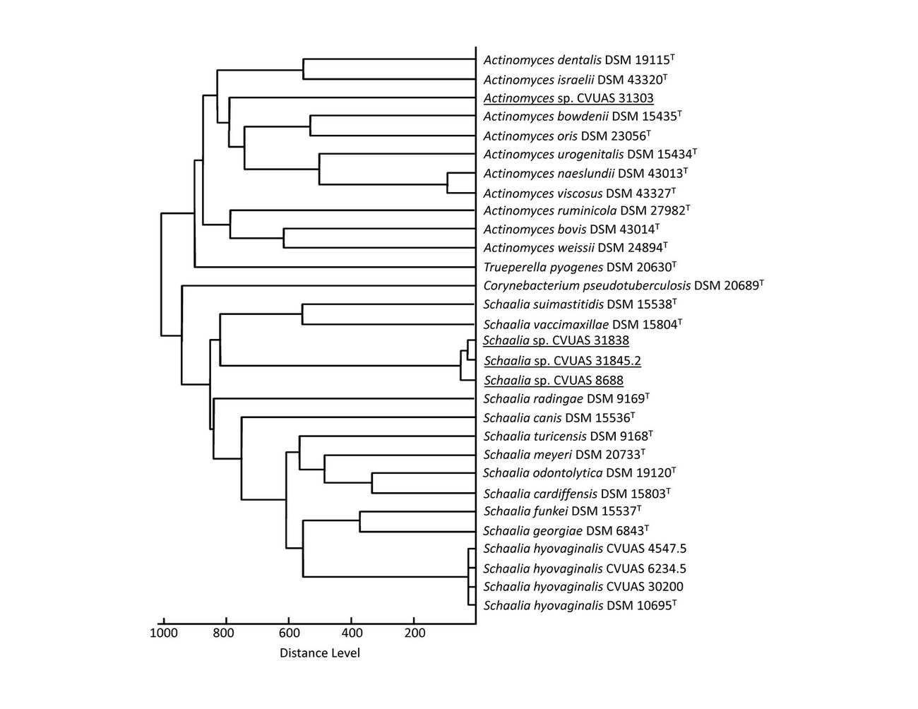
The isolate CVUAS 31303 (goat) could be distinguished from Schaalia isolates using MALDI-TOF MS and was located within the Actinomyces cluster (Fig. 1). The isolates originating from the llama (isolate CVUAS 8688) and the alpacas (isolates CVUAS 31838 and CVUAS 31845.2) exhibit a close relationship clustering on a branch belonging to a separate cluster close to several Schaalia (S.) species, and are therefore referred to as Schaalia species. In contrast, caprine, sheep and porcine S. hyovaginalis field isolates and the S. hyovaginalis type strain DSM 10695, which were identified and verified as S. hyovaginalis by MALDI-TOF MS, are located within a common cluster clearly separated from the isolates originating from the New World camelids (Fig. 1). The MALDI TOF mass-spectra of the isolates and several reference strains are available by exchange via the MALDI-TOF user platform (Rau et al. 2016b).
16S rRNA gene sequencing
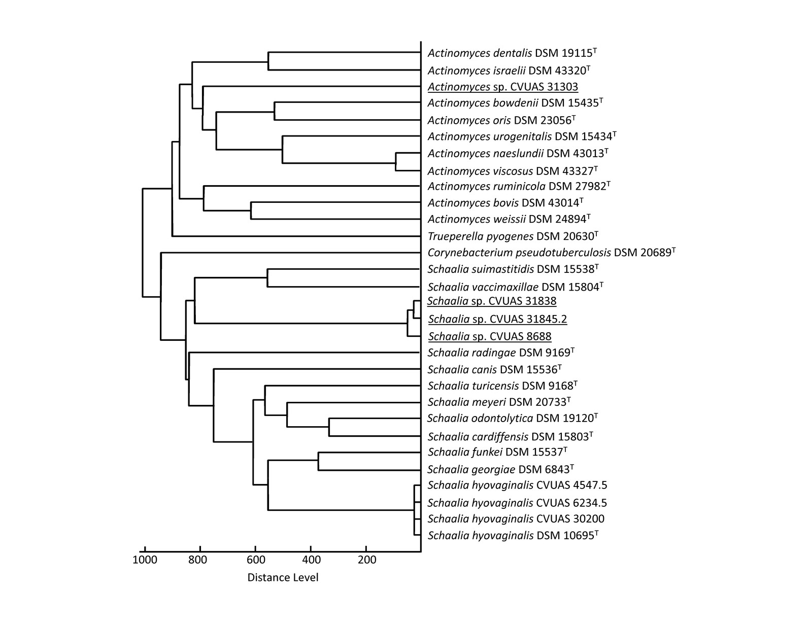
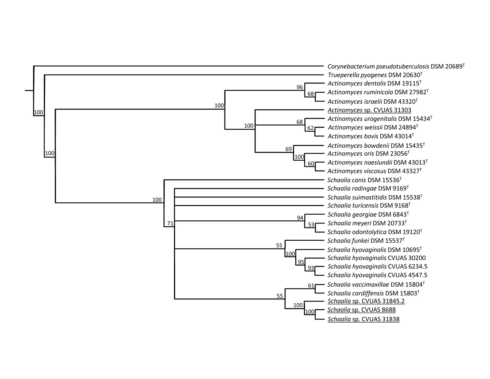
In the 16S rDNA dendrogam, the goat isolate CVUAS 31303 showed a close relationship to Actinomyces species, including Actinomyces bovis. In contrast, the llama and alpaca isolates showed a close relationship in a common sub-cluster within a Schaalia cluster, but were clearly distinct from the cluster comprising sheep, caprine and porcine S. hyovaginalis field isolates including the type strain DSM 10695 (Fig. 2).
Discussion
A considerable proportion of abscess formation in small ruminants and camelids is caused by bacteria belonging to the phylum Actinobacteria. A prominent member of the order Actinomycetales is Corynebacterium (C.) pseudotuberculosis, the causative agent of CLA, which is highly relevant in small ruminants and camelids worldwide (Al-Harbi 2011, Al-Tuffyli and Shekhan 2012, de la Fuente et al. 2017, de Lima e Silva et al. 2016). Control programmes have been implemented in numerous countries worldwide to effectively combat CLA in small ruminants and camelids. Therefore, abscess formation resembling CLA has to be verified by identification of the pathogenic agent, which must be reliably distinguished from C. pseudotuberculosis. Numereous actinomycetes have been described as causative agents of suppurative lesions in humans and animals (Nouioui et al. 2018, Yassin 2014). Among the genera within the family Actinomycetaceae, the recently described novel genus Schaalia has been separated from the genus Actinomyces and reported in connection with abscess formation in humans and various animal species. Case reports have been published on abscess formation due to Actinomyces and Schaalia infections in different localisations such as the brain, subcutis or lymph nodes of goats (Alssahen et al. 2020, Hirai et al. 2007, Ndegwa et al. 2001, Oyekunle et al. 2010, Schumacher et al. 2009), New World camelids (Brown 2006, Fowler 1996), sheep (Alssahen et al. 2020, Collins et al. 2001, Foster et al. 2012), pigs (Hommez et al. 1991, Reichel and Wragg 2007), horses (Chung et al. 2018, Fielding et al. 2008), dogs (Hoyles et al. 2000, Song et al. 2015) and captive and free-ranging wildlife (Alssahen et al. 2020, Gamble and Clancy 2013, Wickhorst et al. 2017). The microorganisms reported in this study, which had been isolated from abscesses in a goat, a llama and two alpacas, attracted our attention because of their similarity to Actinomyces spp. and Schaalia spp., respectively. Further studies on these isolates using MALDI-TOF MS and 16S rDNA analysis initially yielded inconclusive results at species level, despite comprehensive databases that are available for evaluation procedures. However, the isolates could be assigned to bacteria of the genera Actinomyces (goat isolate) and Schaalia (llama and alpaca isolates). The isolate originating from the abscess in the goat is located within the MALDI-TOF MS and 16S rDNA Actinomyces cluster, but is separate to other concrete Actinomyces spp. In contrast, the isolates obtained from the llama and the alpacas show very close relationships to each other in a separate branch next to other Schaalia spp. in both the MALDI-TOF MS and 16S rDNA dendrogram. The close relationship of these actinomycetes in New World camelids, which were isolated at different times and places, suggest the presence of a novel host-adapted Schaalia species. Fowler (1996) also reported on abscess formation in the throat region, lungs or liver and dental abscesses caused by Actinomyces sp. unique to New World camelids and provisionally called these isolates Actinomyces (lamae). The author pointed out that these bacterial isolates may be under-reported due to their morphological appearance as gram-positive, short rods which may be mistaken for cocci.
It is notable that the actinomycetes growth reported in this study was accompanied by F. necrophorum and P. heparinolytica (goat) or T. pyogenes and B. trehalosi (llama). However, no accompanying bacterial aerobic or anaerobic flora could be detected in the abscesses from the two alpacas. Other researchers also report growth of actinomycetes in pure cultures (Brown 2006, Chung et al. 2018, Foster et al. 2012, Oyekunle et al. 2010, Song et al. 2015) or growth in mixed cultures including T. pyogenes, Fusobacterium spp., Bacteroides spp. and Prevotella spp. or Staphylococcus spp. and Streptococcus spp. (Al-Harbi 2011, Collins et al. 2001, Foster et al. 2012, de la Fuente et al. 2017, Gamble and Clancy 2013, Roeder et al. 1989, Schumacher et al. 2009, Wickhorst et al. 2017).
The abscess wounds in the two alpacas were successfully treated by rinsing with iodine solution without additional antibiotic treatment. Unfortunately, no data is available on
the treatment of the abscesses in the goat and the llama. MALDI-TOF MS and 16S rRNA gene analyses suggest that the actinomycetes described in this study represent undesignated novel Actinomyces and Schaalia species. However, this assumption has to be proven by further comprehensive investigations to elucidate the taxonomic status of these interesting, obviously pathogenic actinomycetes.
Conclusion
Actinomycetes represent important bacteria associated with abscess formation. This report shows that so far undescribed actinomycetes are also responsible for abscess formation in addition to already described pyogenic bacteria. Precise and reliable identification at species level is therefore crucial with special reference to CLA (pseudotuberculosis), which is controlled in large-scale control programmess in many countries. Reliable identification of Actinomyces and Schaalia species and detection of undescribed species can be achieved by MALDI-TOF MS and 16S rDNA analyses.
The present study extends our current knowledge about novel actinomycetes associated with abscess formation in small ruminants and New World camelids.
Acknowledgement
The authors thank Jana Ade and Mandy Hailer for performance and evaluation of the PCR assays. We would also like to thank Martin Dyk for carrying out the MALDI-TOF MS analyses and the evaluation of the spectra.
Conflict of interest statement
None of the authors has any financial or personal relationships that could inappropriately influence or bias the content of this paper.
Address for correspondence
Dr. Reinhard Sting
Chemisches und Veterinäruntersuchungsamt Stuttgart
Schaflandstr. 3/3
70736 Fellbach
Germany
reinhard.sting@cvuas.bwl.de
References
Al-Harbi KB (2011): Prevalence and etiology of abscess disease of sheep and goats at Qassim region, Saudi Arabia. Veterinary World 4, 495–499.
Al-Tuffyli YIK, Shekhan MI (2012): Clinical and bacteriological study of subcutaneous abscesses caused by gram positive bacteria in cow and sheep in Al-Qadissiyia province. AL-Qadisiyah Journal of Vet Med Sci 11: 80–85.
Alssahen M, Hassan AA, Rau J, Sammra O, Wickhorst J-P, Lämmler C, Prenger-Berninghoff E, Eisenberg T, Abdulmawjood A (2020): Comparative studies on Schaalia (Actinomyces) hyovaginalis isolated from wild boar, goat and sheep. Berl Münch Tierärztl Wochenschr Online first: 31.03.2020. http://vetline.de/facharchiv/158/3222
Braga WU, Chavera A, Gonzalez A (2006): Corynebacterium pseudotuberculosis infection in highland alpacas (Lama pacos) in Peru. Vet Rec 159: 23–24.
Brown RA (2006): Unusual findings in a llama. Vet Rec 159: 755–756.
Chung ELT, Adamu L, Jesse FFA, Wakil YK, Solomon EM, Turaki UA (2018): Suspected neonatal isoerythrolysis with concurrent Actinomyces hyovaginalis in a foal. J Adv Vet Anim Res 5: 233-239.
Collins MD, Hutson RA, Hoyles L, Falsen E, Nikolaitchouk N, Foster G (2001): Streptococcus ovis sp. nov., isolated from sheep. Int J Syst Evol Microbiol 51: 1147–1150.
Contzen M, Sting R, Blazey B, Rau J (2011): Corynebacterium ulcerans from diseased wild boars carrying Corynebacterium diphteriae-like tox genes. Zoonoses Public Health 58: 479–488.
Fielding CL, Magdesian KG, Morgan RA, Ruby RE, Sprayberry KA (2008): Actinomyces species as a cause of abscesses in nine horses. Vet Rec 162: 18–20.
Foster G, Wragg P, Koylass MS, Whatmore AM, Hoyles L (2012): Isolation of Actinomyces hyovaginalis from sheep and comparison with isolates obtained from pigs. Vet Microbiol 157: 471–475.
Fowler ME (1996): Husbandry and diseases of camelids. Scientific and Technical Review of the International Office of Epizootics 15: 55–169.
de la Fuente R, de las Herasa M, Torrijosa C, Diez de Tejadaa P, Pérez-Sanchob M, Carrióna FJ, Ordena JA, Dominguez-Bernala G (2017): Short communication: Isolation frequency of bacteria causing lymphadenitis and abscesses in small ruminants in central Spain. Small Ruminant Res 154: 5–8.
de Lima e Silva WE, Veneroni Gouveia G, da Conceição Aquino de Sá M., de Simoni Gouveia JJ, de MoraesPeixoto R, Riet-Correa F, Veschi JLA, da Costa MM (2016): Bacteria isolated from abscesses of small ruminants inspected in the semiarid region of Brazil. J Semina Ciênc Agrár 37: 1337–1344.
Gamble KC, Clancy MM (2013): Infectious diseases of concern to captive and free ranging animals in North America. 2nd ed. Infectious Disease Committee, American Association of Zoo Veterinarians, Yulee, Florida
Hirai T, Nunoya T, Azuma R (2007): Actinomycosis of the brain and temporal bone in a goat. J Vet Med Sci 69: 641–643.
Hoyles L, Falsen E, Foster G, Pascual C, Greko C, Collins MD (2000): Actinomyces canis sp. nov., isolated from dogs. Int J Syst Evol 50 Pt 4: 1547–1551.
Hommez J, Devriese LA, Miry C, Castryck F (1991): Characterization of 2 groups of Actinomyces-like bacteria isolated from purulent lesions in pigs. Zentralbl Veterinarmed B 38: 575–580.
Johnson JL (1994): Similarity analysis of rRNAs. In: Methods for General and Molecular Bacteriology, 2nd ed. American Society for Microbiology, Washington DC.
Ndegwa EN, Muleib CM, Munyua SJM (2001): Prevalence of microorganisms associated with udder infections in dairy goats on small-scale farms in Kenya. J S Afr Vet Assoc 72: 97–98.
Nouioui I, Carro L, Garcia-Lopez M, Meier-Kolthoff JP, Woyke T, Kyrpides NC, Pukall R, Klenk HP, Goodfellow M, Goker M (2018): Genome-based taxonomic classification of the phylum Actinobacteria. Front Microbiol 9: 2007.
Oyekunle MA, Talabi AO, Agbaje M, Oni OO, Adebayo AO, Olude MA, Oyewusi IK, Akinduti PA (2010): Actinomycosis in West African dwarf goat in Nigeria. Niger Vet J 31: 80–86.
Pranada BP, Schwarz G, Kostrzewa M (2016): MALDI Biotyping for microorganism identification in clinical microbiology. In: Cramer R (ed) Advances in MALDI and Laser-induced soft ionization mass spectrometry, 1st ed. Springer International Publishing, Basel.
Pruitt K, Brown G, Tatusova T, Maglott D (2002): The Reference Sequence (RefSeq) Database (Updated 2012 Apr 6) In: McEntyre J, Ostell J (eds.). The NCBI Handbook [Internet]. Bethesda (MD). National Center for Biotechnology Information (US), Chapter 18.
Rau J, Eisenberg T, Männig A, Wind C, Lasch P, Sting R (2016a): MALDI-TOF mass spectrometry for reliable identification of bacteria – A validation based on Staphylococcaceae field isolates. Aspects of Food Control and Animal Health (eJournal)2016-03: 1–46. https://ejournal.cvuas.de/issue201603.asp
Rau J, Eisenberg T, Sting R (2016b): MALDI-UP – an internet platform for the exchange of MALDI-TOF mass spectra (eJournal). 2016-01: 1–17. https://ejournal.cvuas.de/issue201601.asp
Reichel R, Wragg P (2007): Isolation of Actinomyces hyovaginalis from a lung lesion in a pig. Vet Rec 160: 203.
Roeder BL, Chengappa MM, Lechtenberg KF, Nagaraja TG, Varga GA (1989): Fusobacterium necrophorum and Actinomyces pyogenes associated facial and mandibular abscesses in blue duiker. J Wildl Dis 25: 370–377.
Schumacher VL, Hinckley L, Gilbert K, Risatti GR, Londoño AS, Smyth JA (2009): Actinomyces hyovaginalis-associated lymphadenitis in a Nubian goat. J Vet Diagn Invest 21: 380–384.
Smith T (1918): A pleomorphic bacillus from pneumotic lungs of calves simulating Actinomyces. J Exp Med 28: 333–344.
Song, RB, Vitullo, CA, da Costa, RC, Daniels, JB (2015): Long-term survival in a dog with meningoencephalitis and epidural abscessation due to Actinomyces species. J Vet Diagn Invest 27: 552–557.
Sprake P, Gold JR (2012): Corynebacterium pseudotuberculosis liver abscess in a mature alpaca (Lama pacos). Can Vet J 53: 387–390.
Wickhorst J, Sammra O, Hassan A, Alssahen M, Lämmler C, Riße K, Eisenberg T, Schauerte N, Geiger C, Prenger-Berninghoff E, Timke M, Abdulmawjood A (2017): Actinomyces hyovaginalis associated with chronic suppurative arthritis in an adult giraffe (Giraffa cameloperdalis reticulate). Berl Münch Tierärztl Wochenschr 130: 161–164.
Yassin AAF (2014): The Family Actinomycetaceae. In: The Prokaryotes, 4th ed. Springer, Berlin Heidelberg.
Zhao K, Li W, Kang C, Du L, Huang T, Zhang X, Wu M, Yue B (2014): Phylogenomics and evolutionary dynamics of the family Actinomycetaceae. Genome Biol Evol 6: 2625–2633.


Kostenfreier Download
Klicken Sie hier, wenn Sie das PDF BMTW-10.23761439-0299-2020-6-Sting.pdf (0.23 MB) herunterladen möchten
Kostenfreier Download
Klicken Sie hier, wenn Sie das PDF BMTW-10.23761439-0299-2020-6-Sting-Tabelle1.pdf (0.12 MB) herunterladen möchten

