Intrathoracic extrapleural infiltrative lipoma in a White Shepherd Dog
Kleintierpraxis 68, 228-235
DOI: 10.2377/0023-2076-XX-228
© M. & H. Schaper GmbH. 2023
Eingereicht: 20. Juni 2022
Akzeptiert: 28. Dezember 2022
Publiziert: 05/2023
Zusammenfassung
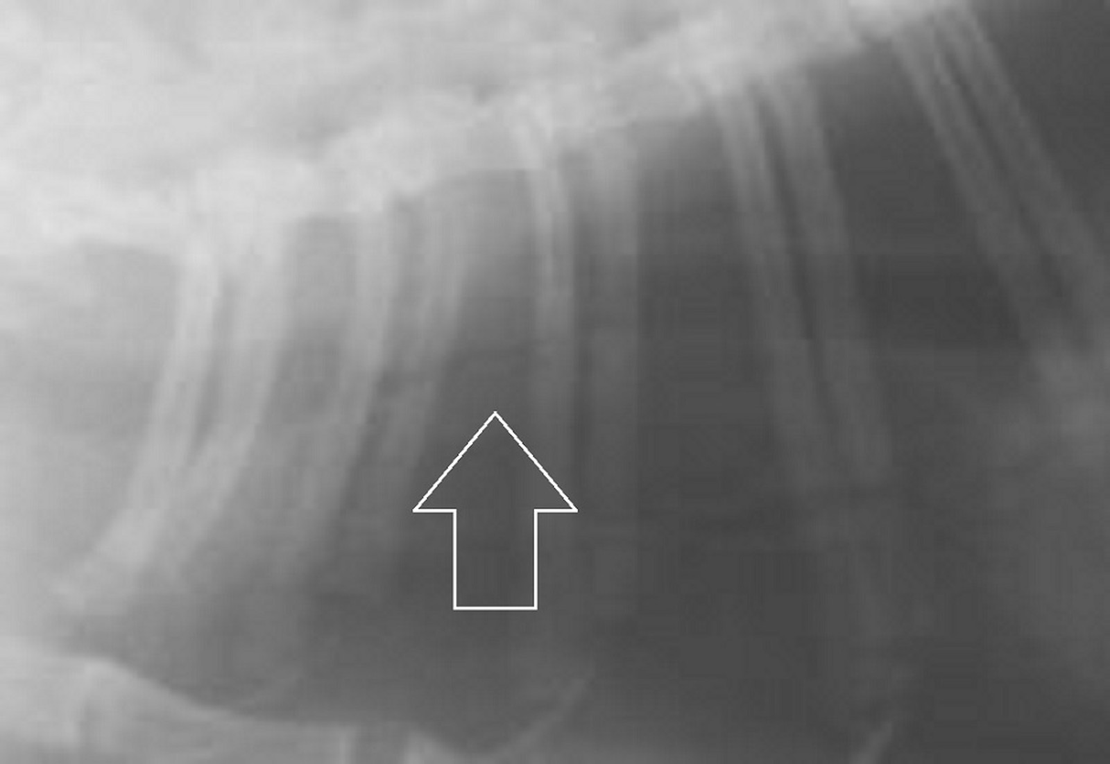
Dieser Fallbericht beschreibt eine achtjährige, unkastrierte, weiße Schäferhündin, die mit Hustensymptomatik vorgestellt wurde, nachdem eine laut Besitzer ungefähr faustgroße, weiche, subkutane Umfangsvermehrung an der rechten seitlichen Brustwand nicht mehr zu palpieren war. Über röntgenologische sowie computertomografische Untersuchungen konnte eine von extra- nach intrathorakal ziehende, fettattenuierende Zubildung dargestellt werden, welche anteilig die Interkostalmuskulatur infiltrierte, intrathorakal Anteile der Lunge nach lateral und über Druck auf das Zwerchfell auch Anteile der Leber nach kaudal verdrängte. Die Masse wurde nach marginaler Resektion mit Rippenteilentfernung und anschließender pathohistologischer Untersuchung als infiltratives Lipom mit gutartiger Fettgewebsnekrose und reparativer Pannikulitis der Thoraxwand identifiziert. Es ist davon auszugehen, dass sich nach Infiltration der Interkostalmuskulatur das Lipom nach intrathorakal und extrapleural fortsetzte, während der extrathorakale, subkutane Anteil größtenteils einem Gewebsuntergang unterlag.
Summary
This case report describes an eight-year-old intact female White Shepherd Dog, which was presented with cough symptoms, after, according to the owner, an about fist-sized, soft, subcutaneous mass on the right chest wall was no longer palpable. X-ray and computed tomographic examination confirmed the presence of a fat-attenuating mass which was invading the thoracic cavity from extra- to intrathoracic, displacing parts of the lung, liver, and partially infiltrating the intercostal muscles. After marginal resection with partial resection of a rib and subsequent pathohistological examination the mass was identified as an infiltrative lipoma with benign adipose tissue necrosis and reparative panniculitis of the thoracic wall. It can be assumed, that the lipoma progressed with an intrathoracic growth after infiltration of the intercostal muscles, while the extrathoracic, subcutaneous part was subject of necrosis.