
Nachweis von FPV, CPV-2a, CPV-2b und FCoV bei Katzen mit Symptomen der felinen Panleukopenie
Berliner und Münchener Tierärztliche Wochenschrift 135, 1–6
DOI: 10.2376/1439-0299-2022-13
© Schlütersche Fachmedien GmbH. 2022
Eingereicht: 11. Juli 2022
Akzeptiert: 27. August 2022
Publiziert: 10/2022
Summary
In the period from August 2019 to August 2020, a total of nine cases of feline panleukopenia (FPV) were detected in two interrelated animal shelter facilities located in the district of Brezno, central Slovakia. All symptomatic cats were tested for the presence of parvovirus in the faeces using a rapid test with all tests being positive. The described animals ranged in age from three months to four years. Post-mortem examinations were performed on six dead animals and samples were taken to determine the presence of feline panleukopenia (FPV), canine parvovirus (CPV) variants 2a and 2b and feline coronavirus (FCoV) in the faeces using the quantitative polymerase chain reaction (qPCR). The examination of the samples was carried out at the Idexx Laboratiories in Leipzig, Germany. Despite intensive therapy, eight cats died and only one survived. This represents a treatment success rate of 11,2%. The findings of the qPCR tests showed positive results for FPV, CPV variants 2a and 2b and for FCoV in the faeces.
These results suggest that co-infection with CPV-2 strains, FCoV and FPV in cats may significantly reduce the success rate of therapy for feline panleukopenia.
Zusammenfassung
Im Zeitraum von August 2019 bis August 2020 wurden in zwei in Relation stehenden Zuchtbetrieben im Bezirk Brezno in der Mittelslowakei insgesamt neun Fälle der felinen Panleukopenie (FPV) festgestellt. Alle symptomatischen Katzen wurden mit einem Schnelltest auf das Vorhandensein des Parvovirus im Kot untersucht, wobei alle betroffenen Tiere positiv getestet wurden. Die beschriebenen Tiere waren im Alter zwischen drei Monaten und vier Jahren. Bei sechs verendeten Tieren wurde eine Obduktion durchgeführt und es wurden Proben entnommen, um das Vorhandensein des felinen Panleukopenievirus (FPV), der Varianten des caninen Parvovirus (CPV) 2a und 2b sowie des felinen Coronavirus (FCoV) mittels der quantitativen PCR-Methode (qPCR) im Kot festzustellen. Die Untersuchungen wurden in den Idexx Labors in Leipzig durchgeführt. Trotz intensiver Therapie starben acht Katzen und nur eine überlebte. Das entspricht einer Erfolgsquote der Behandlung von 11,2 %. Die Ergebnisse der Untersuchungen mittels qPCR waren positiv auf das Vorhandensein von FPV, CPV-2a und b und FCoV im Kot.
Diese Ergebnisse deuten darauf hin, dass eine Koinfektion mit CPV-2-Stämmen, FCoV und FPV bei Katzen die Erfolgsrate der Therapie bei feliner Panleukopenie erheblich verringern kann.
Introduction
Feline panleukopenia is an infectious viral disease caused by members of the genus of Canine protoparvovirus 1, which includes strains of feline panleukopenia virus (FPV) and canine parvovirus 2 (CPV-2) (Allison et al. 2013, 2014). The detection of parvovirus DNA sequences in various carnivore species confirms that parvoviruses have circulated in the population of carnivores for millions of years (Liu et al. 2011). Feline panleukopenia is the oldest known viral disease of the cat. The viral cause of this disease was confirmed in 1928, and the first successful vaccination against feline panleukopenia was performed in 1934 (Verge and Cristoforoni 1928, Leasure et al. 1934). The CPV-2 and FPV parvoviruses are so closely related that their NS1 protein is at least 85% identical. A significant difference between them is that feline parvovirus is relatively genetically stable and undergoes genetic changes only very slowly, whereas the opposite is true in the case of CPV-2 (Shackelton et al. 2005, Hoelzer et al. 2008, Cotmore et al. 2014). Canine parvovirus 2 probably evolved from feline parvovirus through changes in five or six amino acid positions in the capsid protein or from another closely related parvovirus (Shackelton et al. 2005). In the first half of the 1980s, CPV-2 evolved into two variants (CPV-2a and CPV-2b). The antigenic variant of CPV-2a was recognized in 1984 and differs in the antigenic epitope in the substitution in VP2, the replacement of the Asn residue at position 426 by Asp and the replacement of Ile at position 555 by Val (Parrish et al. 1985). The most recent discovery of a new subtype was made in Italy in 2000. This is the third variant of canine parvovirus type 2 (CPV-2c). CPV-2c differs from CPV-2b in a single amino acid residue at position 426 where Asp has been replaced by Glu. The Glu mutation at position 426 affects the major antigenic region located above the three-fold spike of the CPV-2 capsid (Buonavoglia et al. 2001). Interestingly, CPV-2 has long been unable to induce infection in cats. However, with further adaptation to canids, changes in amino acid positions resulted in CPV-2 binding better to canid cellular receptors, but also having the ability to infect cats (Hueffer and Parrish 2003). Thus, newer CPV-2 strains of CPV-2a, CPV-2b and CPV-2c have acquired a new ability to infect cats. Furthermore, they may cause a disease in cats with clinical signs similar to feline panleukopenia (Truyen et al. 1995, Mochizuki et al. 1996, Truyen et al. 1996). The potential pathogenicity of CPV-2 strains in cats, however, is still unclear. Experimental infections of cats with CPV-2a/b strains suggest that these strains have lower pathogenicity in cats and may induce persistent infection (Battilani et al. 2006). In our study, we describe co-infection with CPV-2a, CPV-2b, FPV and FCoV in the faeces of animals in which hypothermia, apathy and vomiting occurred and low efficacy of therapy was observed.
Material and methods
Patient details
The age of the affected animals ranged from three months to four years (mean age was 16.2 months). None of the patients were neutered. There were six males and three females among the described patients. The patients came from two different facilities; six patients were living in private keeping with access to the outside environment and three cats came from a shelter. All cats were of the European shorthair cat breed. None of the patients had ever been vaccinated against FPV before, and all patients were regularly treated against endoparasites.
Case history
The presented patients were referred to our veterinary outpatient clinic for treatment of mild apathy, intermittent vomiting and anorexia between August 2019 and August 2020. Six cats were brought for treatment on the second day after the onset of clinical signs of disease, and three patients on the fourth day after the first signs of disease.
Clinical examination
After admission, the patients underwent clinical examinations, including a measurement of the body temperature, an assessment of the condition of the oral mucosa and conjunctivae, an auscultatory examination of the chest, and palpation of the abdominal cavity and peripheral lymph nodes. Hydration status was also assessed in each patient.
Laboratory examination
After clinical examinations, blood was collected from the Vena cephalica antebrachii of the cats for haematological and biochemical examinations and rectal swabs were taken to detect the presence of parvovirus in the faeces using the CPV Ag Test Kit (BioNote Inc., South Korea). Haematological and biochemical blood tests were carried out in the laboratories of Unilabs Slovakia, s.r.o. Six animals were also autopsied, and their faecal samples were collected to investigate the presence of FPV, CPV-2a, CPV-2b and FCoV by identifying the parvovirus using the quantitative PCR method, which facilitates distinguishing mutations on the VP2 gene. PCR tests were performed at the IDEXX laboratories in Leipzig, Germany, using the LightCycler® 480 system (Roche, Mannheim, Germany).
Therapy
All patients received causal therapy with immunoglobulins, fluid therapy, antibiotic therapy to cover the anaerobic and gram-negative spectrum, symptomatic therapy to prevent vomiting, and supportive therapy focusing on artificial feeding and application of vitamin preparations (Greene 2012).
The results
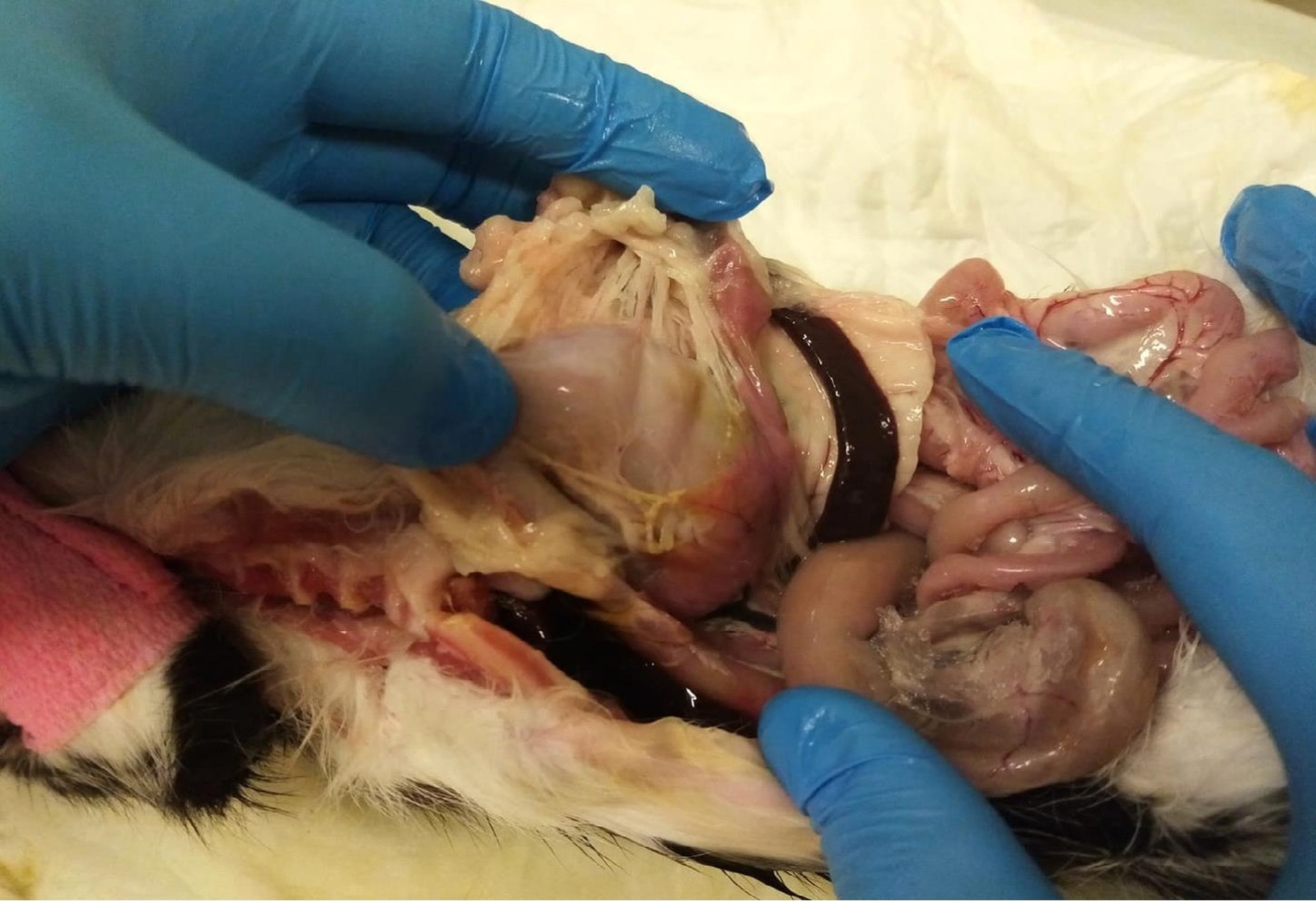
In the affected animals, mild apathy (in seven patients), anorexia (in eight patients), intermittent vomiting (in eight patients) and marked hyperthermia (in nine patients, up to 41°C) were the predominant symptoms at initial treatment. Diarrhoea was not present in any of the animals. The results of the rapid tests for parvovirus in the faeces were positive in all animals. For clarity, the results of the haematological and biochemical blood tests are only presented for the animals in which the presence of FCoV, FPV, CPV-2a and CPV-2b was detected by qPCR. Haematological examinations of the blood revealed leukopenia in five animals (the mean of 3,67 x 10⁹/l, the reference range of 6,00–11,00) and leucocytosis in one animal (16,94 x 10⁹/l). Thrombocytopenia was found in five animals (the mean of 62,83 x 10⁹/l, the reference range of 180,00–500,00) while a normal platelet count was found in one animal. Biochemical examinations of the blood revealed elevated levels of aspartate aminotransferase (AST) in six animals (the mean of 3,32 μcat/l, the reference range of 0,00–0,56). PCR results were positive for FPV, CPV-2a and b, and for FCoV. The results of haematological and AST tests in cats with the PCR detection of FPV, CPV-2a, CPV-2b and FCoV are shown in Table 1. Out of the nine cases presented, eight patients died and one cat survived, representing a success rate of 11,2%. The only surviving animal was a 4-year-old male cat. At post-mortem examination, four cats were found to have non-specific findings without gastroenteritis. One cat had pancreatitis without gastroenteritis and one cat was found to have suppurative nephritis and enteritis (Fig. 1 and 2).
Discussion
Feline panleukopenia is a severe infectious disease affecting cats of different ages. It is characterized by high morbidity and mortality, causing severe damage to the mucosa of the small intestine, leading to severe enteritis with subsequent diarrhoea and dehydration associated with destruction of leukocytes and lymphocytes in the lymph nodes (Gaskell et al. 1996, Esfandiari and Klingeborn 2000, Simpson and Birnbaum 2006). In some cases, genetic recombination between CPV-2 and FPV isolates could be observed. About 95% of cases of feline panleukopenia tend to be caused by FPV and about 5% by CPV-2 strains (Greene 2012). For instance, in Germany, CPV-2 virus has been detected in only 10% of affected cats, whereas in Southeast Asia it has been proven in up to 80% of affected cats (Stuetzer and Hartmann 2014). According to a study conducted by Byrne et al. (2018) in Australia, CPV-2 was not detected in a single faecal sample among samples collected from from 218 female and male cats from animal shelters. In contrast, other authors have detected CPV-2 strains in faeces of clinically healthy animals using PCR and virus isolation (Mochizuki et al. 1993, Clegg et al. 2012, Mukhopadhyay et al. 2017). The pathogenicity of CPV-2 variants in cats has not been fully elucidated. Some studies suggest that CPV- 2 has the same pathogenic potential as FPV in cats (Mochizuki et al. 1996, Truyen et al. 1996, Decaro et al. 2010, Battillani et al. 2011); in other studies, no clinical signs were observed in infected animals except transient leukopenia (Chalmers et al. 1999, Nakamura et al. 2001). These results have led to speculation that CPV-2 may be more likely to cause asymptomatic and persistent infection in cats compared to FPV, although further studies are needed to fully understand the potential of cats as carriers of CPV-2. The study by Balboni et al. (2018) found an equal prevalence of FPV and CPV-2 in the samples examined, but these samples were predominantly from asymptomatic cats. In general, CPV-2 is not a common cause of feline panleukopenia, although only a small number of cats showing signs of feline panleukopenia were tested for the presence of CPV-2. Natural CPV-2 infection in cats showing signs of panleukopenia has been described in several cats (Mochizuki et al. 1996, Decaro et al. 2010, Miranda et al. 2014). In 1 case, a fatal co-infection with FPV and CPV-2a was described in a 3-month-old Persian cat (Battilani et al. 2013). According to Green (2012), clinical signs of feline panleukopenia tend to be milder in infection with CPV-2a and CPV-2b strains compared to the infection caused by FPV. Co-infections of FPV and CPV-2 have been described in several cats (Battilani et al. 2011, Battilani et al. 2013). Li et al. (2018) demonstrated co-circulation of FPV with a novel CPV-2a and of CPV-2 virus strains in cats with signs of gastroenteritis in China. The aim of our study was to present cases of feline panleukopenia with clinical signs consistent with feline panleukopenia but with a slightly different disease course and findings of FPV, CPV-2a, CPV-2b and FCoV, in which we observed low treatment efficacy and high mortality. According to Sherding (1989), the success rate is around 75% if the therapy is chosen correctly. However, according to other retrospective studies, the therapy success rate ranges from 20% to 51% (Kruse et al. 2011, Litster and Benjanirut 2014, Porporato et al. 2018). Barrs (2019) reports mortality rates of 50% to 80% despite therapy, and Isaya et al. (2021) reported the mortality rate 42,9%. That said, in our case series, the success rate was only 11,2 % despite intensive therapy, which is a very low value compared to the studies described. It is possible that such a high mortality rate was caused by co-infection with FPV and CPV-2 strains alone. However, it is also possible that the mortality rate was significantly increased by the presence of FCoV. For example, in dogs, coronavirus infection has a low mortality rate, but co-infection with CPV-2 is known to significantly increase mortality rate in dogs with coronavirus (Alves et al. 2018). Another possible cause of the high mortality in the presented case series is the FPV strain itself (antigenic variant) responsible for the infection, as changes in the VP2 protein affect the antigenicity and host range of FPV and CPV-2 (Truyen et al. 1995, Allison et al. 2013). Another factor that could increase the mortality rate in the presented cases is secondary bacterial infection, despite the fact that all patients received antibiotic therapy to suppress anaerobic and gram-negative bacteria. Further studies are needed to determine the prevalence of CPV-2 strains in cats with clinical signs of feline panleukopenia and to compare mortality rates associated with co-infection with FPV and FCoV.
Conclusion
In the case series of feline panleukopenia with low treatment efficacy that we presented, we found the canine parvovirus strains CPV-2a and CPV-2b in addition to FPV and FcoV in six samples examined. This finding is interesting because only a small number of cats affected with feline panleukopenia globally were tested for the presence of canine parvovirus CPV-2 and its strains CPV-2a, CPV-2b and CPV-2c simultaneously with the detection of FPV and FCoV. This is the first study in Slovakia to look at the detection of CPV-2a and CPV-2b strains in cats affected with panleukopenia and co-infection with FPV, FCoV and CPV-2b strains. Future studies with larger numbers of affected cats are needed to determine the percentage of canine parvovirus strains in cats with panleukopenia and to analyse the course of the disease and the efficacy of therapy in these patients.
Conflict of interest
The authors declare that they have no proprietary, professional or other personal interest in any product or company that may influence the content or opinions expressed in this publication.
Acknowledgement
This publication is the result of the implementation of the project: „Open scientific community for modern interdisciplinary research in medicine (OPENMED)”, ITMS2014+: 313011V455 supported by the Operational Programme Integrated Infrastructure, funded by the ERDF. This work was supported by the Ministry of Education, Science, Research and Sport of the Slovak Republic through the project KEGA 008UVLF-4/2022.
Ethical approval
The study was performed in compliance with the institutional guidelines for animal welfare issued by The ethical committee of the University of Veterinary Medicine and Pharmacy in Košice. Written informed consents were obtained from all of the patient owners.
Authors contribution
Conceptualization: AC, JM.
Methodology: AC, JM, BV.
Investigation and data curation: AC, MD, LZ.
Writing – original draft: AC.
Writing – review & editing: AC, JM, BV, MD, LZ.
All authors have read and approved the final manuscript.
Address for correspondence
Monika Drážovská
Department of Epizootiology, Parasitology and Protection of One Health
University of Veterinary Medicine and Pharmacy in Košice
Komenského 73, 041 81 Slovakia
monika.drazovska@uvlf.sk
Literature
Allison AB, Kohler DJ, Fox KA, Brown JD, Gerhold RW, Shearn-Boschler VI, Dubovi EJ, Parrish CR, Holmes EC (2013): Frequent Cross-Species Transmission of Parvoviruses among Diverse Carnivore Hosts. J Virol 87(4): 2342–2347.
Allison AB, Kohler DJ, Ortega A, Hoover EA, Grove DM, Holmes EC, Parrish CR (2014): Host-specific parvovirus evolution in nature is recapitulated by in vitro adaptation to different carnivore species. PLoS Pathogens 10(12): e1004586.
Alves C, Granados OFO, Budaszewski RDF, Streck AF, Weber MN, Cibulski SP, Pinto LD, Ikuta N, Canal CW (2018): Identification of enteric viruses circulating in a dog population with low vaccine coverage. Brazilian J Microbiol 49: 790–794.
Balboni A, Bassi F, Arcangeli S, Zobba R, Dedola C, Alberti A, Battilani M (2018): Molecular analysis of carnivore Protoparvovirus detected in white blood cells of naturally infected cats. BMC Vet Res 14: 41.
Barrs VR (2019): Feline panleukopenia: a re-emergent disease. Vet Clin North Am Small Anim Pract 49(4): 651–670.
Battilani M, Scagliarini A, Ciulli S, Morganti L, Prosperi S (2006): Hight genetic diversity of the VP2 gene of a canine parvovirus strain detected in a domestic cat. Virology 352(1): 22–26.
Battilani M, Balboni A, Ustulin M, Giunti M, Scagliarini A, Prosperi S (2011): Genetic complexity and multiple infections with more Parvovirus species in naturally infected cats. Vet Res 42(1): 43.
Battilani M, Balboni A, Giunti M, Prosperi S (2013): Co-infection with feline and canine parvovirus in a cat. Vet Ital 49(1): 127–129.
Buonavoglia CV, Martella A, Pratelli M, Tempesta A, Cavalli D, Bozzo G, Decaro N, Carmichael LE (2001): Evidence for evolution of canine parvovirus type-2 in Italy. J Gen Virol 82: 1555–1560.
Byrne P, Beatty JA, Šlapeta J, Corley SW, Lyons RE, McMichael L, Kyaw-Tanner MT, Dung PT, Decaro N, Meers J, Barrs VR (2018): Shelter-housed cats show no evidence of faecal shedding of canine parvovirus DNA. Vet J 239: 54–58.
Chalmers WSK, Truyen U, Greenwood NM, Baxendale W (1999): Efficacy of feline panleucopenia vaccine to prevent infection with an isolate of CPV2b obtained from a cat. Vet Microbiol 69(1-2): 41–45.
Clegg SR, Coyne KP, Dawson S, Spibey N, Gaskell RM, Radford AD (2012): Canine parvovirus in asymptomatic feline carriers. Vet Microbiol 157(1-2): 78–85.
Cotmore SF, Agbandje-McKenna M, Chiorini JA, Mukha DV, Pintel DJ, Qiu J, Soderlund-Venermo M, Tattersall P, Tijssen P, Gatherer D, Davison AJ (2014): The family Parvoviridae . Arch Virol 159: 1239–1247.
Decaro N, Buonavoglia D, Desario C, Amorisco F, Colaianni ML, Parisi A, Terio V, Elia G, Lucente MS, Cavalli A, Martella V, Buonavoglia C (2010): Characterisation of canine parvovirus strains isolated from cats with feline panleukopenia. Res Vet Sci 89(2): 275–278.
Esfandiari J, Klingeborn B (2000): A comparative study of a new rapid and one‐step test for the detection of parvovirus in faeces from dogs, cats and mink. J Vet Med 47(2): 145–153.
Gaskell RM, Tennant B, Bennett M, Willoughby K (1996): Feline and Canine Infectious Diseases. Iowa State Press, Ames, IA.
Greene CE (2012): Canine Viral Enteritis Infections Diseases of the Dog and Cat. 4th ed. Saunders Elsevier, St. Louis.
Hoelzer K, Shackelton LA, Holmes EC, Parrish CR (2008): Within-host genetic diversity of endemic and emerging parvoviruses of dogs and cats. J Virol 82(22): 11096–11105.
Hueffer K, Parrish CR (2003): Parvovirus host range, cell tropism and evolution. Curr Opin Microbiol 6: 392–398.
Isaya R, Ciccarelli S, Enache D, Specchi S, Pesaresi M, Ferri F, Porporato F, Auriemma E, B, Coppola LM, Zini E (2021): Correction to: Gastrointestinal ultrasonographic findings in cats with Feline panleukopenia: a case series. BMC Vet Res 17(1): 143.
Kruse BD, Unterer S, Horlacher K, Sauter-Louis C, Hartmann K (2011): Feline Panleukopenie – differierender Krankheitsverlauf bei Katzen im Alter von unter bzw. über 6 Monaten. Tierarztl Prax Kleintiere 39(04): 237–242.
Leasure EE, Lienardt HF, Taberner FR (1934): Feline infectious enteritis. North Am Vet 15: 30–34.
Li X, Wu H, Wang L, Spilbey N, Liu C, Ding H, Liu W, Liu Y, Tian K (2018): Genetic characterization of parvoviruses in domestic cats in Henan province, China. Transbound Emerg Dis 65(6): 1429–1435.
Litster A, Benjanirut CH (2014): Case series of feline panleukopenia virus in an animal shelter. J Feline Med Surg 16(4): 346–353.
Liu H, Fu Y, Xie J, Cheng J, Ghabrial SA, Li G, Peng Y, Yi X, Jiang D (2011): Widespread Endogenization of Densoviruses and Parvoviruses in Animal and Human Genomes. J Virol 85(19): 9863–9876.
Miranda C, Parrish CR, Thompson G (2014): Canine parvovirus 2c infection in a cat with severe clinical disease. J Vet Diagn Invest 26(3): 462–464.
Mochizuki M, Harasawa R, Nakatani H (1993): Antigenic and genomic variabilities among recently prevalent parvoviruses of canine and feline origin in Japan. Vet Microbiol 38(1-2): 1–10.
Mochizuki M, Horiuchi M, Hiragi H, San Gabriel MC, Yasuda N, Uno T (1996): Isolation of canine parvovirus from a cat manifesting clinical signs of feline panleukopenia. J Clin Microbiol 34: 2101–2105.
Mukhopadhyay HK, Nookala M, Hangamani NR, Sivaprakasam A, Antony PX, Thanislass J, Srinivas MV, Pillai RM (2017): Molecular characterisation of parvoviruses from domestic cats reveals emergence of newer variants in India. J Feline Med Surg 19(8): 846–852.
Nakamura K, Ikeda Y, Miyazawa T, Tohya Y, Takahashi E, Mochizuki M (2001): Characterisation of cross-reactivity of virus neutralising antibodies induced by feline panleukopenia virus and canine parvoviruses. Res Vet Sci 71(3): 219–222.
Parrish CR, O’Connell PH, Evermann JF, Carmichael LE (1985): Natural variation of canine parvovirus. Science 230: 1046–1048.
Porporato F, Horzinek MC, Hofmann-Lehmann R, Ferri F, Gerardi G, Contiero B, Vezzosi T, Rocchi P, Auriemma E, Lutz H, Zini E (2018): Survival estimates and outcome predictors for shelter cats with feline panleukopenia virus infection. J Am Vet Med Assoc 253(2): 188–195.
Shackelton LA, Parrish CR, Truyen U, Holmes EC (2005): High rate of viral evolution associated with the emergence of carnivore parvovirus. Proc Nat Acad Sci USA 102: 379–384.
Sherding RG (1989): Small bowel disease. In: Ettinger SJ (ed.), Textbook of Veterinary Medicine. 3rd ed. WB Saunders, Philadelphia, 1351–1353.
Simpson KW, Birnbaum N (2006): Fluid and electrolyte disturbances in gastrointestinal and pancreatic disease. In: Dibartola SP (ed.), Fluid, Electrolyte and Acid-Base Disorders in Small Animal Practice. 3rd ed. Elsevier Saunders, St. Louis, MO, 420–436.
Stuetzer B, Hartmann K (2014): Feline parvovirus infection and associated diseases. Vet J 201: 150–155.
Truyen U, Gruenberg A, Chang SF, Veijalainen P, Obermaier B, Parrish CR (1995): Evolution of the feline subgroup parvoviruses and the control of canine host range. J Virol 69: 4702–4710.
Truyen U, Evermann JF, Vickler E, Parrish CR (1996): Evolution of canine parvovirus involved loss and gain of the feline host range. Virology 215: 186–189.
Verge J, Cristoforoni N (1928): La gastroenterite infectieuse des chats estelle due a un virus filtrable? Comptes Rendus Seances Biologies 99: 312–314.


Kostenfreier Download
Klicken Sie hier, wenn Sie das PDF BMTW-10.23761439-0299-2022-13-Drážovská.pdf (0.3 MB) herunterladen möchten
Kostenfreier Download
Klicken Sie hier, wenn Sie das PDF BMTW-10.23761439-0299-2022-13-Drážovská-Tabelle1.pdf (0.05 MB) herunterladen möchten