
Spontane Lipidembolie bei einem adipösen Minipig (Sus scrofa domesticus)
Berliner und Münchener Tierärztliche Wochenschrift 136, 1–4
DOI: 10.2376/1439-0299-2022-20
© Schlütersche Fachmedien GmbH. 2023
Eingereicht: 4. Oktober 2022
Akzeptiert: 19. Januar 2023
Publiziert: 02/2023
Summary
A perished, one-year-old, male-neutered minipig was submitted for pathological examination after a brief history of anorexia and hypothermia. Necropsy revealed an obese body condition and multifocal to coalescing severe necroses of the visceral fatty tissue, especially of the perirenal region. Histopathologic examination showed severe chronic granulomatous and necrotising steatitis and optically empty vacuoles in the lumen of small and capillary blood vessels of lung, liver and kidney, consisting of lipid droplets ascertained by Sudan red stain. Lipid embolism occurs after traumatic insults such as long-bone fracture or orthopaedic surgery in both human and veterinary medicine. Non-traumatic lipid embolism, as observed in this case, is rare and the pathogenesis is still obscure.
Zusammenfassung
Ein verendetes, einjähriges, männlich-kastriertes Minischwein wurde nach eintägiger Inappetenz und Untertemperatur zur pathologischen Untersuchung eingesandt. In der Sektion zeigte sich der Tierkörper in adipösem Ernährungszustand und wies hochgradige, multifokale bis konfluierende Nekrosen des viszeralen Fettgewebes auf, die im perirenalen Bereich besonders ausgeprägt waren. In der histologischen Untersuchung zeigten sich eine hochgradige, chronische granulomatös-nekrotisierende Steatitis und in Lunge, Leber und Nieren optisch leere Vakuolen in den Lumina von kleinkalibrigen und kapillären Gefäßen. Mittels Sudanrot-Färbung stellten sich diese Vakuolen als Fetttröpfchen dar. Lipidembolien treten im Zusammenhang mit traumatischen Insulten, beispielsweise Knochenbrüchen oder Operationen, sowohl in der Human- als auch in der Veterinärmedizin auf. Nicht-traumatische Lipidembolien, wie in diesem Fall, sind selten und die Pathogenese ist immer noch unklar.
Introduction
Lipid emboli are lipid droplets entering the blood circulation and leading to a partial or complete clogging of small vessels and capillaries. In human medicine, the clinical manifestation of lipid embolism is called “fat embolism syndrome” (FES) characterised by respiratory distress, neurological signs and cutaneous petechiae (Milroy und Parai 2019). Fat embolism is a frequent and critical complication of long bone fractures or orthopaedic surgeries, particularly those featuring traumatisation of medullary contents (Hulman 1995). In men, lipid embolism is also uncommonly reported unrelated to traumatic insults (Shapiro und Hayes 1984). The prevalence ranges from less than 1% to 29% depending on clinical criteria alone (<1%) or with taking account of pathological findings (up to 29%) (Fabian et al. 1990, Bulger et al. 1997, Shaikh 2009). The non-traumatic form occurs in association with fatty liver disease (Sakashita et al. 2017), hyperlipidaemia and hypertension (Meng et al. 2020), bone marrow necrosis with haemoglobinopathies (Gangaraju et al. 2016), acute pancreatitis (Lynch 1954), corticosteroid therapy, lymphography and fat emulsion therapy (Shaikh 2009). In veterinary medicine, reports of spontaneous non-traumatic lipid embolism are even more uncommon. The mechanisms underlying non-traumatic lipid embolism are still obscure. The following report presents a case of a privately kept minipig with a spontaneous fatal lipid embolism.
Case report
Animal history
Top Job:
A one-year-old male-neutered minipig showed a short period of anorexia, a poor general condition and hypothermia with a body temperature of 34°C. According to the owner, the minipig died with a short outcry. The flock consisted of two minipigs and two domestic pigs. The feed consisted of pig pellets for final fattening.
Pathologic and histologic examination
The body condition of the minipig was obese with a bodyweight of 86 kg. The visceral fat tissue showed severe, multifocal to coalescing, firm, pale yellow, friable masses interpreted as fat necroses, which were most prominent at the perirenal region. The lung was moderately retracted and showed severe, diffuse alveolar oedema, moderate, diffuse alveolar emphysema and moderate, diffuse congestion. Moderate to severe diffuse congestion was further detected in the liver, kidneys and meninges. Grossly, all long bones, the vertebral column and the pelvis showed after longitudinal cutting no evidence of bone fractures. Pancreas and thyroid glands displayed a normal anatomic appearance and subcutaneous haemorrhages suggestive of a traumatic impact were not found.
A broad spectrum of tissues and organs was collected and fixed in 10% neutral buffered formalin for 24 hours, followed by trimming and embedding in paraffin wax. Haematoxylin and eosin (H&E) stained slides were prepared by standard histotechnological procedures for microscopic examination.
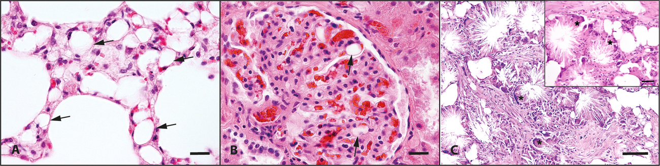
Histologically in the lung, sharply demarcated, optically empty vacuoles were present in the lumen of small-calibre blood vessels expanding the alveolar septa (Fig. 1A). Additionally, there were mild to moderate alveolar histiocytosis and slight hyperplasia of type II pneumocytes. Lower numbers of similar vacuoles were also detected in renal glomerular capillaries (Fig. 1B) and small hepatic blood vessels. The macroscopically altered visceral adipose tissue showed extensive areas of ruptured adipocytes with complete loss of cytological details and deposition of numerous cholesterol needles. The necrotic tissue was surrounded and infiltrated with numerous macrophages and multinucleated giant cells (foreign body type). Areas of necrosis were separated by strands of fibroblastic connective tissue (Fig. 1C). In addition, a multifocal mild, lympho-histiocytic myocarditis, epicarditis and endocarditis was detected. The myocardium also showed a mild, multifocal lipomatosis cordis. The intestinal wall was severely thickened by deposition of adipose tissue within the submucosa and between the inner, circular and outer, longitudinal musculature layers. Histological examination of pancreas, thyroid gland and bone marrow of sternum and femur revealed no morphological changes.
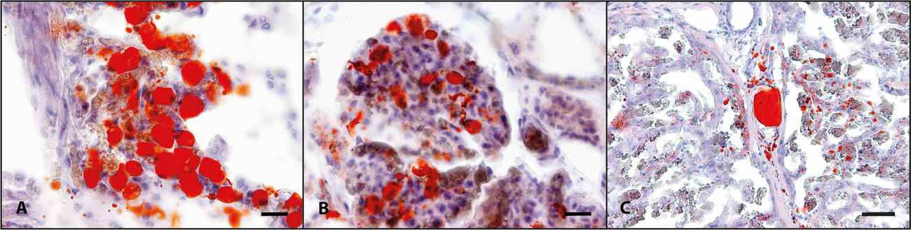
To further characterise the vacuoles in lung, liver and kidneys, Sudan-III red staining was applied on cryosections resulting in the identification of intravascular lipid vacuoles in lung, kidneys (Fig. 2A, 2B), and liver (Fig. 2C).
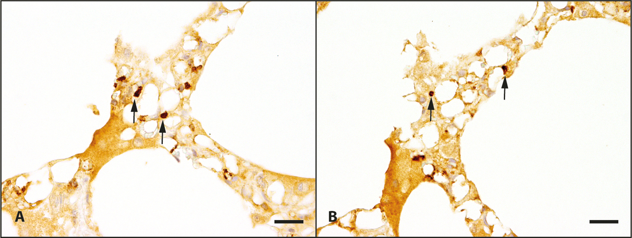
Using antibodies against CD31 and factor VIII-related antigen, both immunohistochemical markers for vascular endothelial cells, and the avidin-biotin-peroxidase complex method (Junginger et al. 2012), immunohistochemistry revealed that the vacuoles were located in vascular structures lined by endothelial cells (Fig. 3A, 3B).
Conclusion
According to the pathological examination, the minipig died of an acute cardiac and circulatory failure due to systemic lipid embolism. A traumatic cause for the lipid embolism, such as a bone fracture, was not detected. Therefore, a spontaneous, idiopathic lipid embolism has to be considered, whilst other predisposing diseases including pancreatic, thyroidal or bone marrow disorders, were not found. Spontaneous fatal lipid embolism is rare in man and is assumed to be underdiagnosed. Reports in veterinary medicine are even more uncommon. Case reports include a horse with yellow fat disease (Suárez-Bonnet et al. 2008), a dog with diabetes mellitus (Brown und Fitzpatrick 1986), a Vietnamese potbellied pig (Newkirk et al. 2012) and cohorts of minipigs used for obesity studies (Renner et al. 2018, Viuff et al. 2020). According to the latter reports, all pigs showed an obese body condition and severe, chronic visceral fat necrosis suggesting a possible causal relationship of metabolic factors associated with obesity and chronic fat necrosis in the development of lipid embolism.
The pathogenesis of lipid embolism is still unclear. Two theories have been set up, the mechanical and the biochemical theory. According to the mechanical theory, in traumatic cases, lipid components from ruptured adipocytes, for example from the bone marrow or adipose tissue, presumably enter the blood stream directly. The biochemical theory implies that metabolic factors in combination with inflammatory components may play a more significant role in the pathogenesis of lipid embolism in non-traumatic cases (Hulman 1988). In vitro studies showed that fat emulsion composed of very low density lipoproteins (VLDL) and chylomicrons aggregates to form macroglobules in presence of C-reactive protein (CRP) and calcium (Rowe et al. 1986). Thus, elevated plasma lipid levels caused by underlying metabolic conditions, in combination with pro-inflammatory factors such as elevated CRP, might lead to the aggregation of lipid components in vivo, with possible subsequent blockage of small blood vessels and capillaries (Hulman 1995).
In the present case, the obese body condition, the fat necroses as well as the inappetence shortly before death, are potential factors that might have caused elevated plasma lipid levels through an increased mobilisation of free fatty acids (FFA). In minipigs, both obesity and severe inflammation of the visceral fat tissue can cause elevated CRP levels (Renner et al. 2018, Viuff et al. 2020). Although plasma lipid levels and CRP were not measured in vivo, lipid embolism in the present case was most likely caused by a combination of metabolic and inflammatory factors.
The cause of the necrosis with associated chronic granulomatous inflammation of the adipose tissue is not known. One possible impact may be an insufficient angiogenesis of excessive adipose tissue in obese animals resulting in tissue compression by its weight with subsequent hypoxia and necrosis (Schwarz et al. 2000). Another driver for necrosis and inflammation of fatty tissue may be a deficiency of vitamin E, well described in pigs and horses as yellow fat disease (Nafstad und Tollersrud 1970, Suárez-Bonnet et al. 2008). A deficiency of vitamin E results in peroxidation of unsaturated fatty acids resulting in degenerative changes to many different tissues and cells, and is mostly associated with myocardial and skeletal muscle degeneration and liver necrosis. Changes affecting fatty tissue, visceral as well as subcutaneous, are brown-yellow discoloration caused by ceroid and lipofuscin deposition within macrophages and adipocytes (Suárez-Bonnet et al. 2008). Details of the animals´ feed and nutritional status are not available, but the lack of associated lesions, the distribution of the necrosis and the lack of ceroid pigment within affected fat tissue make a vitamin E deficiency unlikely in the present case.
In cases with histologically empty vacuoles in lumen of blood vessels, air embolism should also be considered as a differential diagnosis especially with limited patient´s history. Using Sudan-III red staining it was shown that the intravascular vacuoles contained lipid components and air embolism was ruled out.
In conclusion, this report represents a rare case of spontaneous lipid embolism that is probably related to obesity and fat necrosis. Previous reports of lipid embolism in minipigs support this association and it should be considered that fatal lipid embolism may be a possible consequence of obesity in minipigs.
Acknowledgement
The authors thank the staff of the Department of Pathology, University of Veterinary Medicine Hannover, for the excellent technical support.
Conflict of interest
The authors have no conflicts of interest related to the case described.
Ethical Approval
The authors hereby declare that they have followed the universally accepted guidelines of good scientific practice while preparing the present paper.
Financing
This Open Access publication was funded by the Deutsche Forschungsgemeinschaft (DFG, German Research Foundation) - 491094227 „Open Access Publication Costs“ and the University of Veterinary Medicine Hannover, Foundation.
Author Contribution
Necropsy, macroscopic and histologic examination and drafting of manuscript: SF.
Clinical examination, critical revision of article: AvA.
Histologic examination, interpretation and critical revision of article: WB.
Macroscopic and histologic examination and interpretation, critical revision of article: JJ.
Histologic examination and interpretation, critical revision of article and approval of final draft for publication: PW.
Address for correspondence
Sandra Franke Institut für Pathologie Stiftung Tierärztliche Hochschule Hannover Bünteweg 17 30559 Hannover Sandra.Franke@tiho-hannover.de
Literatur
Brown TP, Fitzpatrick RK (1986): Glomerular lipid emboli in a diabetic dog. Vet Pathol 23: 209–211.
Bulger EM, Smith DG, Maier RV, Jurkovich GJ (1997): Fat embolism syndrome. A 10-year review. Arch Surg 132: 435–439.
Fabian TC, Hoots AV, Stanford DS, Patterson CR, Mangiante EC (1990): Fat embolism syndrome: prospective evaluation in 92 fracture patients. Crit Care Med 18: 42–46.
Gangaraju R, Reddy VV, Marques MB (2016): Fat embolism syndrome secondary to bone marrow necrosis in patients with hemoglobinopathies. South Med J 109: 549–553.
Hulman G (1995): The pathogenesis of fat embolism. J Pathol 176: 3–9.
Hulman G (1988): Pathogenesis of non-traumatic fat embolism. Lancet 1: 1366–1367.
Junginger J, Schwittlick U, Lemensieck F, Nolte I, Hewicker-Trautwein M (2012): Immunohistochemical investigation of Foxp3 expression in the intestine in healthy and diseased dogs. Vet Res 43: 23.
Lynch MJ (1954): Nephrosis and fat embolism in acute hemorrhagic pancreatitis. AMA Arch Intern Med 94: 709–717.
Meng Y, Zhang M, Ling H, Huang S, Miao Q, Yu Y, Zhang F, Qiu P, Li D (2020): Nontraumatic multiple-organ fat embolism: an autopsy case and review of literature. Am J Forensic Med Pathol 41: 131–134.
Milroy CM, Parai JL (2019): Fat embolism, fat embolism syndrome and the autopsy. Acad Forensic Pathol 9: 136–154.
Nafstad I, Tollersrud S (1970): The vitamin E-deficiency syndrome in pigs. I. Pathological changes. Acta Vet Scand 11: 452–480.
Newkirk KM, Fineschi V, Kiefer VR, VanAmstel SR (2012): Lipid emboli in a Vietnamese potbellied pig (Sus scrofa). J Vet Diagn Invest 24: 625–629.
Renner S, Blutke A, Dobenecker B, Dhom G, Müller TD, Finan B, Clemmensen C, Bernau M, Novak I, Rathkolb B, Senf S, Zöls S, Roth M, Götz A, Hofmann SM, Hrabĕ de Angelis M, Wanke R, Kienzle E, Scholz AM, DiMarchi R, Ritzmann M, Tschöp MH, Wolf E (2018): Metabolic syndrome and extensive adipose tissue inflammation in morbidly obese Göttingen minipigs. Mol Metab 16: 180–190.
Rowe IR, Soutar AK, Pepys MB (1986): Agglutination of intravenous lipid emulsion (‚Intralipid‘) and plasma lipoproteins by C-reactive protein. Clin Exp Immunol 66: 241–247.
Sakashita M, Sakashita S, Sakata A, Uesugi N, Ishige K, Hyodo I, Noguchi M (2017): An autopsy case of non-traumatic fat embolism syndrome. Pathol Int 67: 477–482.
Schwarz T, Morandi F, Gnudi G, Wisner E, Paterson C, Sullivan M, Johnston P (2000): Nodular fat necrosis in the feline and canine abdomen. Vet Radiol Ultrasound 41: 335–339.
Shaikh N (2009): Emergency management of fat embolism syndrome. J Emerg Trauma Shock 2: 29–33.
Shapiro MP, Hayes JA (1984): Fat embolism in sickle cell disease. Report of a case with brief review of the literature. Arch Intern Med 144: 181–182.
Suárez-Bonnet A, Espinosa de los Monteros A, Herráez P, Rodríguez F, Andrada M, Caballero MJ (2008): Fat embolism secondary to yellow fat disease in an Appaloosa horse. J Vet Diagn Invest 20: 684–687.
Viuff BM, Straarup EM, Nowak J, Morgills L, Skydsgaard M, Sjögren I, Wulff BS, Christoffersen B (2020): Lipid embolism in obese Göttingen minipigs. Toxicol Pathol 48: 379–392.

Kostenfreier Download
Klicken Sie hier, wenn Sie das PDF BMTW-10.2376-1439-0299-2022-20-Franke.pdf (0.4 MB) herunterladen möchten