

Prolaps von Umfangsvermehrungen des Geschlechtstrakts aus der Vaginalöffnung als Vorstellungsgrund weiblicher Meerschweinchen (Cavia porcellus)
Kleintierpraxis 65, 4–11
DOI: 10.2377/0023-2076-65-4
© M. & H. Schaper GmbH. 2020
Publiziert: 01/2020
Summary
Pathological changes affecting the female reproductive tract in guinea pigs have been described previously in the literature. However, knowledge about the clinical findings and management of uterine and vaginal proliferative diseases has been sparse to date. In dogs, a common clinical presentation of various types of uterine and vaginal abnormalities is a tumor-like mass protruding from the vaginal orifice. In the present study, we investigated the frequency and underlying causes of masses protruding from the vaginal orifice as the reason for presentation in guinea pigs. A retrospective analysis of pathological and associated clinical records of a total of 723 intact female guinea pigs identified masses protruding from the vaginal orifice in 12 animals (1.7%). Of these, six originated from the vagina – four vaginal leiomyomas and two non-neoplastic vaginal polyps– and six from the uterus – two cervical gland neoplasms, two decidual cell proliferations, and two uterine prolapses. Additionally, four cases of decidual cell proliferations being discharged from the uterus and subsequently found in the animal’s cage were identified. All six vaginal masses were successfully removed by simple ligation through the vaginal opening. The resection of three uterine masses by simple ligation resulted in an incomplete surgical removal. In conclusion, differential diagnoses for vulvar masses in guinea pigs should include vaginal or uterine neoplastic or non-neoplastic proliferative disorders. Knowledge of the anatomical origin and type of disease is essential for the therapeutic management. Most notably, although decidual cell proliferations are thought to regress spontaneously, they may also be associated with uterine neoplasia and so necessitate a thorough clinical examination of the uterus.
Zusammenfassung
Erkrankungen des Geschlechtstrakts weiblicher Meerschweinchen sind in der aktuellen Literatur gut beschrieben. Dennoch ist das Wissen über klinische Befunde und die Behandlung uteriner und vaginaler Veränderungen nur begrenzt. Beim Hund sind Vorfälle uteriner oder vaginaler Massen aus der Vaginalöffnung ein durchaus bekannter Vorstellungsgrund. In der vorliegenden Studie wurden daher Häufigkeit und Art pathologischer Veränderungen, die mit einem Gewebsprolaps aus der Vaginalöffnung verbunden sind, beim Meerschweinchen untersucht.
Hierfür wurden in einer retrospektiven Analyse die klinischen und pathologischen Befunde von insgesamt 723 weiblich-intakten Meerschweinchen gesichtet. Bei zwölf dieser Tiere (1,7 %) konnte ein Gewebeprolaps aus der Vaginalöffnung nachgewiesen werden. Sechs dieser Veränderungen waren vaginalen Ursprungs – vier vaginale Leiomyome und zwei nicht-neoplastische, vaginale Polypen –, während die anderen sechs dem Uterus entstammten – zwei Neoplasien der zervikalen Drüsen, zwei deziduale Proliferationen und zwei Uterusvorfälle. Zusätzlich wurden vier Fälle dezidualer Proliferationen festgestellt, welche ausgeschieden und anschließend vom Besitzer im Käfig gefunden wurden. Alle vaginalen Massen konnten durch die Vaginalöffnung mittels einfacher Ligatur erfolgreich entfernt werden. Die Entfernung drei uteriner Massen mittels einfacher Ligatur hatte eine unvollständige Resektion zur Folge.
Die Ergebnisse der vorliegenden Studie weisen darauf hin, dass bei Gewebsvorfällen aus der Vaginalöffnung sowohl vaginale als auch uterine neoplastische und nicht-neoplastische, proliferative Veränderungen differenzialdiagnostisch berücksichtigt werden sollten. Eine Untersuchung des anatomischen Ursprungs und der Art der Veränderung trägt maßgeblich zur Entscheidung bei, welche Therapie gewählt werden sollte. Bemerkenswerterweise wird bei dezidualen Proliferationen von einer spontanen Regression ausgegangen. Sie können jedoch ebenfalls mit uterinen Neoplasien assoziiert sein. Daher wird eine gründliche klinische Untersuchung des Uterus angeraten.
Introduction
Retrospective pathological studies have identified several types of uterine and vaginal masses in pet guinea pigs (Bertram et al. 2018b, Laik-Schandelmaier et al. 2017, Sommerey et al. 2004, Veiga-Parga et al. 2016). However, information regarding the etiology and frequency of vulvar masses is sparse (Eatwell 2003). In dogs, a very common clinical presentation of a variety of vaginal and uterine disorders is a mass protruding from the vaginal orifice (Azari et al. 2010, Manothaiudom and Johnston 1991, Miller et al. 2008, Sontas et al. 2010, Suzuki et al. 2006, Thacher and Bradley 1983). In contrast, masses protruding from the vaginal orifice have only rarely been described in rodents (e.g. a vaginal leiomyoma in a chinchilla; Bertram et al. 2018a). In guinea pigs, only anecdotal reports of uterine prolapse after parturition exist (Kondert and Mayer 2017). Nevertheless, guinea pigs with masses protruding from the vaginal orifice have been occasionally presented to the authors over the last years. Therefore, the aim of this retrospective case study was to characterize the clinical presentation by describing the anatomical origin and underlying type of disorder as well as gaining first insights into possible effective treatment options.
Material and methods
The aim of the present study was to describe the clinical findings and management as well as the pathological findings of guinea pigs with lesions protruding from the vaginal orifice. As such, all pathological and associated clinical records between 1995 and June 2018 of female intact pet guinea pigs were reviewed for masses protruding from the vaginal orifice. During that period of time, a total of 655 postmortem examinations of female intact pet guinea pigs were performed and 64 surgical biopsies from the female genital tract were examined. A retrospective summary of the pathological findings of these cases, including 73 cases of uterine and six vaginal neoplastic and non-neoplastic masses, has been described previously (Bertram et al. 2018b). Additionally, four masses discharged from the female genital tract and submitted for histopathological examination were included in the present study. The types of disease affecting the cases were all confirmed by histopathology. All the masses, except for uterine prolapse, were further characterized by immunohistochemistry with primary antibodies against vimentin (clone V9), alpha smooth muscle actin (clone ASM-1), desmin, cytokeratin (clones AE1/AE3), estrogen receptors (ER), and, in two cases, also with antibodies against von Willebrand factor (vWF) to exclude vascular neoplasia (Table 1).
Top Job:
Case descriptions
Twelve of the 719 guinea pigs included in the study (1.7%) (mean age: 3 years; range: 2–7 years) had been presented within a few hours after a mass was detected protruding from the vaginal orifice (Table 2). These included six vaginal neoplastic or tumor-like masses, four uterine neoplastic or tumor-like masses, and two non-neoplastic uterine prolapses. Additionally, in four guinea pigs (mean age: 4 years; range: 3.5–7 years), tumor-like masses were discharged from the female genital tract and subsequently found in the home cage by the owners. The following sections will describe the clinical and pathological findings for the different disease types separately.
Vaginal leiomyoma
The clinical examination of cases Nos. 1–4 determined that the masses protruding from the vaginal orifice were pedunculated and connected to the wall inside the vagina. Three of the four masses were removed under general anesthesia using simple ligation and resection at the base of the mass. In case No. 4, the owners declined surgical management and elected for humane euthanasia with subsequent postmortem examination.
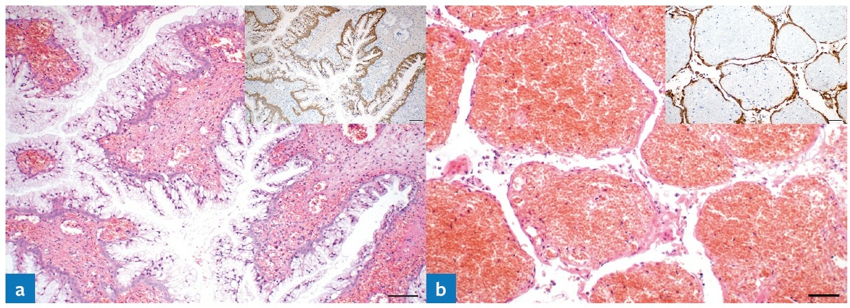
The vaginal masses had a diameter ranging from 1.1 to 2.2 cm. The histopathology of all four masses revealed streams of uniform spindle cells with a single, elongated, and bluntly ending nucleus (Fig. 1a). Mitotic figures were one or less in ten high-power fields (400x magnification). The surgical margins were free of tumor cells in the three cases with surgical removal. In all four cases, the cells stained immunohistochemically positive for vimentin, alpha smooth muscle actin (SMA), desmin (Fig. 1a, inset), and estrogen receptor, but were negative for cytokeratin. Hence, the masses were diagnosed as vaginal leiomyomas. In case No. 4, the post-mortem examination additionally identified bilateral ovarian cysts, moderate glandular-cystic endometrial hyperplasia, and a mild, acute, diffuse, heterophilic endometritis and vaginitis.
Vaginal polyps (tumor-like masses)
In cases No. 5 and 6, the clinical examination revealed that the masses protruding from the vaginal orifice were also pedunculated and connected to the vaginal wall. Both masses were removed under general anesthesia using simple ligation and resection at the base of the mass. They had diameters of 0.9 cm and 1.6 cm, respectively. Histopathologically, the masses contained a regularly arranged epithelial lining and variable amounts of glandular structures entrapped in an abundant fibrovascular stroma (Fig. 1b). In both cases, multifocal epithelial ulceration and moderate, acute, stromal hemorrhage with moderate, heterophilic infiltration were present. The histopathological diagnosis in both cases was non-neoplastic exophytic vaginal polyps.
Endocervical gland neoplasia
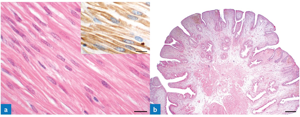
In cases No. 7 and 8, the masses protruding from the vaginal orifice showed severe hemorrhage. The clinical examination (including vaginoscopy) could not determine their exact anatomical origin. Regardless, the masses were resected through the vaginal orifice after simple ligation of its pedicle as basally as possible. After formalin-fixation, the masses had a length of 1.8 cm and 1.5 cm, respectively. Histopathologically, both surgical biopsies revealed tubulopapillary growth, consisting of folds and crypts of mainly single-layered columnar epithelial cells resembling endocervical glands (Fig. 2a). The tumor cells contained abundant, foamy cytoplasm and small, hyperchromatic nuclei. The fibrovascular stroma of both masses was markedly expanded by severe, acute, diffuse hemorrhage in large areas of the section (Fig. 2b). In case No. 8, columnar to flattened tumor cells showed advanced cellular and nuclear pleomorphism with multifocal stratification and squamous metaplasia. In both cases, the tumor cells extended to the surgical margins, i.e. the surgical resection was incomplete. The tumor cells were immunohistochemically positive for cytokeratin (Fig. 2a and 2b, inset) and negative for vimentin, SMA, desmin, and von Willebrand factor (vWF), consistent with epithelial neoplasia. Based on the histopathological and immunohistochemical findings, cases No. 7 and 8 were diagnosed as a cervical gland adenoma and adenocarcinoma, respectively.
Decidual cell proliferations
Cases No. 9 and 10 presented with a brown mass protruding from the vaginal orifice by 2.0 and 3.0 cm, respectively. Guinea pig No. 9 reportedly also showed dyschezia. In this case, both radiographs and ultrasound images suggested a uterine origin and the animal underwent ovariohysterectomy (OHE). Its recovery was uneventful and the animal died four years later due to unrelated reasons. In case No. 10, a vaginal tumor was initially suspected, however, radiographic examination did not reveal the exact origin (Fig. 3). The mass was subsequently resected through the vaginal orifice via simple ligation at its pedicle as basally as possible. Under general anesthesia, the mass was found to be originating from the uterus; however, the owner declined an OHE.
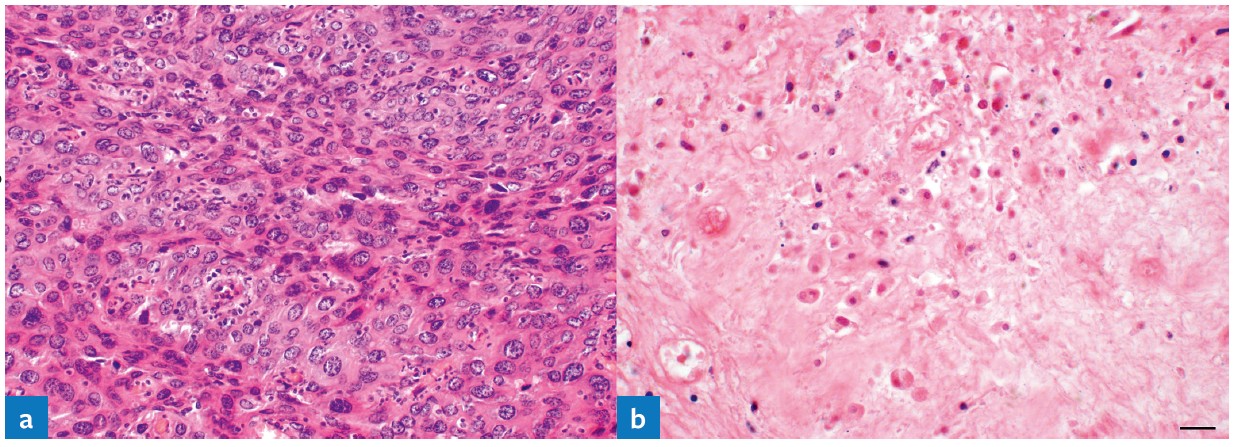
The histopathology of both masses revealed multiple, distinct but intermingling cellular populations (Fig. 4a): large, polygonal, epithelioid cells with abundant eosinophilic cytoplasm and round to oval, hypochromatic nuclei as well as densely-packed, spindle-shaped cells containing oval to elongated nuclei. Moderate cellular and nuclear pleomorphism were observed and some cells were bi- or multinucleated. Distinct regions showed necrosis with multifocal hemorrhage. In case No. 10, the lesion involved the surgical margins. Most cells were immunohistochemically positive for vimentin, SMA, and estrogen receptor in both cases, as well for desmin in case No. 9. Both cases were negative for cytokeratin and case No. 10 additionally for desmin. The masses were diagnosed as decidual cell proliferations which consist of two types: deciduoma or decidualization. Since the entire uterus was submitted for case No. 9, this mass could be further differentiated as a uterine deciduoma.
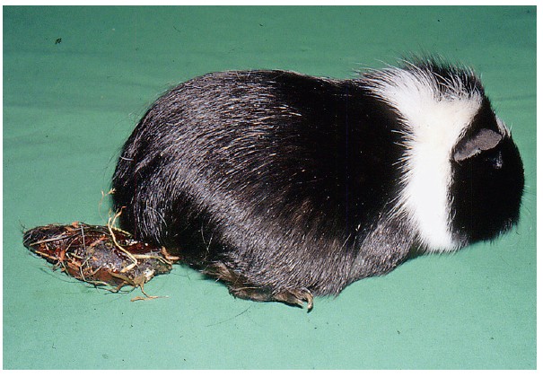
Uterine prolapse
In cases No. 11 and 12, a long, tube-like structure was found in the anogenital region (Fig. 5). In case No. 11, the mass was primarily considered a rectal prolapse. After an unsuccessful attempt of manual repositioning and the owner´s decline of surgical management, the guinea pig was humanely euthanized and submitted for postmortem examination. In case No. 12, a uterine prolapse was clinically suspected and repositioned under general anesthesia with subsequent OHE. The pathological examination identified a uterine prolapse and bilateral cystic rete ovarii in both cases. Glandular-cystic endometrial hyperplasia was additionally diagnosed in case No. 12.
Discharged masses
In cases No. 13 to 16, the owners found a hemorrhagic mass in the cage of their female guinea pigs. The animals were subsequently presented to a veterinarian for clinical examination and the masses subjected to histopathological examination. The owners of all four animals also reported a chronic, hemorrhagic, vaginal discharge previous to the discharge of the mass. The clinical examination and radiography suggested uterine uterine enlargement in two of the four guinea pigs. The histopathology of the masses in all four cases were largely identical to the decidual cell proliferations of cases No. 9 and 10. However, widespread necrosis was a far more dominant feature (Fig. 4b). Immunohistochemically, the four masses were positive for vimentin, SMA, and estrogen receptors and negative for cytokeratin and desmin; similar to case No. 10. These findings were consistent with uterine decidual cell proliferations including uterine deciduoma or decidualization.
Discussion
Based on the present findings, several differential diagnoses including neoplastic and non-neoplastic lesions need to be considered for masses protruding from the genital opening in female guinea pigs. Whilst a protruding mass is a common clinical presentation of guinea pigs with vaginal disorders, uterine disorders, which occur far more frequently, only occasionally protrude through the vaginal orifice. Interestingly, half of the masses in the twelve animals in the present study originated from the uterus and the other half from the vagina. Besides the disorders described in the present study (i.e. vaginal leiomyoma, vaginal polyps, cervical gland neoplasm, uterine prolapse, and decidual cell proliferation), the differential diagnosis should also include fetal or placental retention. It is additionally important to distinguish vaginal protrusions from rectal prolapses.
Furthermore, uterine decidual cell proliferation may be discharged from the genital tract and subsequently found in the animal’s cage. To the author’s knowledge, this has not been reported previously and future studies are required to determine its frequency. Hemorrhagic vaginal discharge had been observed prior to the discovery of the discharged mass. However, a mass protruding from the vaginal orifice or any other location had not been observed by the owners. Nevertheless, histopathology and immunohistochemistry strongly support a uterine origin, consistent with uterine decidual cell proliferation (Dixon et al. 2014, Laik-Schandelmaier et al. 2017, Yoshizawa et al. 2013).
Based on the pathological findings of the discharged decidual cell proliferations, it was, however, not possible to clearly distinguish between deciduoma and focal decidualization. Both lesions originate from uterine decidual stromal cells and show an almost identical histopathological appearance. According to Laik-Schandelmaier et al. (2017), a proliferation attached to the luminal part of a uterine epithelial tumor can be classified as focal superficial decidualization, whilst a solitary change without any underlying pathological lesion is considered a deciduoma. Therefore, thorough uterine examination is required. Both types of decidual cell proliferation represent a non-neoplastic, proliferative response to hormonal stimuli and mechanical irritation or traumatization (Dixon et al. 2014, Mitchell and Garris 1978). Deciduoma have been shown to regress spontaneously in guinea pigs (Mitchell and Garris 1978), which has not been reported for decidualization. However, this type of decidual cell proliferation is generally associated with uterine epithelial neoplasia, such as adenoma and adenocarcinoma (Bertram et al. 2018b, Laik-Schandelmaier et al. 2017) and OHE may be indicated. The underlying cause of detachment of some decidual cell proliferations from the uterus is currently unknown. However, we speculate that this may result from widespread necrosis, as observed in all the decidual cell proliferations in the present study. Further research is required to determine the adequate clinical management of guinea pigs with decidual cell proliferation. Deciduoma probably do not require treatment as they regress spontaneously; however, we strongly recommend a thorough examination of the uterus to exclude uterine neoplasia which is associated with focal decidualization.
The findings of the present study indicate that the differentiation between vaginal and uterine origin of the protruding mass is essential, as different forms of surgical management will be required. Complete removal of the discrete vaginal masses was confirmed via histopathological examination of the surgical margins. As such, ligation and resection through the vaginal orifice was considered adequate treatment for these benign vaginal masses. However, a limitation of the present study is the unavailability of follow-up information. Conversely, invasive, intramural vaginal neoplasia may require more invasive surgical techniques (Nelissen and White 2012). In contrast to vaginal masses, all the uterine masses were incompletely removed when accessed through the vagina. Therefore, uterine masses require OHE. Deciduoma are probably an exception as they tend to regress spontaneously (Mitchell and Garris 1978). However, in all cases of decidual cell proliferation, it should be ensured that the decidual cell proliferation is not associated with uterine neoplasia as discussed above. For diagnostic determination of the origin, various imaging methods such as radiography and ultrasonography may be used (Beregi et al. 2001, Bertram et al. 2018a, Fuchs et al. 2013, Manothaiudom and Johnston 1991, Skoric et al. 2010). Although only performed in one of the present cases, vaginoscopy has been discussed as being a highly appropriate diagnostic method for certain diseases of the genital tract in canine practice (Azari et al. 2010, Kydd and Burnie 1986, Manothaiudom and Johnston 1991, Nelissen and White 2012). The technical requirements and procedures of vaginoscopy have previously been described for guinea pigs and other rodents (Divers 2010, Erlacher-Reid et al. 2012, Lennox 2005). Vaginal cytology cannot only be used for estrus detection but also for the diagnosis of mainly inflammatory disorders (Kondert and Mayer 2017). Whether vaginal cytology may be beneficial for the differentiation of neoplastic and non-neoplastic vaginal and uterine disorders in guinea pigs needs to be shown by future studies.
Conclusion
In guinea pigs, masses of vaginal origin commonly present as protruding from the vaginal orifice. In contrast, although uterine tumors occur more frequently than vaginal masses in this species, they rarely protrude from the vaginal orifice. The differential diagnosis of vaginal masses in guinea pigs should include vaginal leiomyoma, vaginal polyps, cervical gland tumors, uterine decidual cell proliferations (deciduoma or decidualization), and uterine prolapse. It is important to distinguish between vaginal or uterine origin and also between the different types of disorders as they require different types of clinical management, especially regarding the possible surgical approach. For example, deciduoma often do not require surgical management as they tend to regress spontaneously; however, it is important to exclude uterine neoplasia which is associated with uterine decidualization.
Fazit
Vorfälle vaginaler Massen aus der Vaginalöffnung kommen bei weiblichen Meerschweinchen häufiger vor als bisher angenommen. Jedoch können sich gelegentlich auch uterine Veränderungen, die generell häufiger vorkommen, durch einen Vorfall aus der Vaginalöffnung präsentieren. Es sollten unter anderem folgende Differenzialdiagnosen berücksichtigt werden: vaginale Leiomyome, vaginale Polypen, Neoplasien der zervikalen Drüsen, uterine deziduale Proliferationen – Deziduome und Dezidualisationen – und Uterusvorfälle. Ursprung – Vagina oder Uterus – sowie die Art der Veränderungen sollten unterschieden werden, da diese die klinischen Überlegungen, insbesondere im Hinblick auf einen chirurgischen Eingriff, entscheidend beeinflussen können. So wird beispielsweise davon ausgegangen, dass Deziduome nicht chirurgisch entfernt werden müssen, da sie sich in der Regel spontan zurückbilden; jedoch sollte der Uterus genau untersucht werden, um uterine Neoplasien, welche mit Dezidualisationen assoziiert sind, auszuschließen.
Conflict of interest
The authors would like to confirm that they have no protected, financial, occupational or other personal interests in a product, service and/or a company which could influence the content or opinions presented in the manuscript.
Ethical approval
We strongly support the recommendations by COPE (publicationethics.org) and ARRIVE (svg.to/arrive).
Authors contribution
All the authors contributed to the writing, editing and final approval of the manuscript.
Address for correspondence
PD Dr. Kerstin Müller
Klinik für kleine Haustiere
Freie Universität Berlin
Oertzenweg 19b
14163 Berlin
Germany
Kerstin.Mueller@fu-berlin.de
References
Azari O, Akhtardanesh B, Derakhshanfar A, Oloumi MM, Vosough D (2010): Vaginal cavernous hemangioma with cystic ovaries and cystic endometrial hyperplasia in a dog. Comp Clin Pathol 19: 413–416.
Beregi A, Molnár V, Perge E, Felkai C (2001): Radiography and ultrasonography in the diagnosis and treatment of abdominal enlargements in five guinea pigs. J Small Anim Pract 42: 459–463.
Bertram C, Klopfleisch R, Müller K (2018a): Vaginal Leiomyoma in a Chinchilla (Chinchilla laniger). J Small Anim Pract 59: 583.
Bertram C, Müller K, Klopfleisch R (2018b): Genital tract pathology in female pet guinea pigs (Cavia porcellus): a retrospective study of 655 post-mortem and 64 biopsy cases. J Comp Pathol 165: 13–22.
Divers SJ (2010): Exotic mammal diagnostic endoscopy and endosurgery. Vet Clin North Am Exot Anim Pract 13: 255–272.
Dixon D, Alison R, Bach U, Colman K, Foley GL, Harleman JH, Haworth R, Herbert R, Heuser A, Long G (2014): Nonproliferative and proliferative lesions of the rat and mouse female reproductive system. J Toxicol Pathol 27: 1S.
Eatwell K (2003): Ovarian and uterine disease in guinea pigs: a review of five cases. Exotic DVM 5: 37–39.
Erlacher-Reid C, Gallagher A, Brock A, Hall N (2012): Use of rigid endoscopy to evaluate vaginal haemorrhage in a rat. Vet Rec 170: 262.
Fuchs S, Eberhardt F, Niesterok C, Kiefer I (2013): Ultraschall bei Kaninchen und Meerschweinchen – Häufigste pathologische Befunde des Geschlechtsapparats. kleintier konkret 16: 23–28.
Kondert L, Mayer J (2017): Reproductive medicine in guinea pigs, chinchillas and degus. Vet Clin North Am Exot Anim Pract 20: 609–628.
Kydd D, Burnie A (1986): Vaginal neoplasia in the bitch: a review of forty clinical cases. J Small Anim Pract 27: 255–263.
Laik-Schandelmaier C, Klopfleisch R, Schöniger S, Weiffenbach G, Staudacher M, Aupperle H (2017): Spontaneously arising tumours and tumour-like lesions of the cervix and uterus in 83 pet guinea pigs (Cavia porcellus). J Comp Pathol 156: 339–351.
Lennox A (2005): Endoscopy of the distal urogenital tract as an aid in differentiating causes of urogenital bleeding in small mammals. EXOTIC DVM 7: 43–47.
Manothaiudom K, Johnston SD (1991): Approach to vaginal/vestibular masses in the bitch. Vet Clin North Am Small Anim Pract 21: 509–521.
Miller JM, Lambrechts NE, Martin RA, Sponenberg D, Subasic M (2008): Persistent vulvar hemorrhage secondary to vaginal hemangioma in dogs. J Am Anim Hosp Assoc 44: 86–89.
Mitchell JA, Garris D (1978): Deciduoma formation in response to uterine trauma in the guinea pig. Biol Reprod 19: 1135–1140.
Nelissen P, White RA (2012): Subtotal vaginectomy for management of extensive vaginal disease in 11 dogs. Vet Surg 41: 495–500.
Skoric M, Fictum P, Jekl V, Hauptman K, Knotek Z, Hermanova M (2010): Vaginal leiomyosarcoma in a degu (Octodon degus): a case report. Vet Med (Praha) 55: 409–412.
Sommerey C, Köhler K, Reinacher M (2004): Erkrankungen des Meerschweinchens aus Sicht der Pathologie. Tierarztl Prax Ausg K Kleintiere Heimtiere 32: 377–383.
Sontas B, Altun E, Güvenc K, Arun S, Ekici H (2010): Vaginal neurofibroma in a hysterectomized poodle dog. Reprod Domest Anim 45: 1130–1133.
Suzuki K, Nakatani K, Shibuya H, Sato T (2006): Vaginal rhabdomyosarcoma in a dog. Vet Pathol 43: 186–188.
Thacher C, Bradley R (1983): Vulvar and vaginal tumors in the dog: a retrospective study. J Am Vet Med Assoc 183: 690–692.
Veiga-Parga T, La Perle KM, Newman SJ (2016): Spontaneous reproductive pathology in female guinea pigs. J Vet Diagn Invest 28: 656–661.
Yoshizawa K, Emoto Y, Kinoshita Y, Kimura A, Uehara N, Yuri T, Shikata N, Tsubura A (2013): Histopathological and immunohistochemical characterization of spontaneously occurring uterine deciduomas in young adult rats. J Toxicol Pathol 26: 61–66.


Kostenfreier Download
Klicken Sie hier, wenn Sie das PDF KTP-2020-01-Bertram.pdf (0.38 MB) herunterladen möchten
Kostenfreier Download
Klicken Sie hier, wenn Sie das PDF KTP-2020-01-Bertram-Tabelle1.pdf (0.03 MB) herunterladen möchten
Kostenfreier Download
Klicken Sie hier, wenn Sie das PDF KTP-2020-01-Bertram-Tabelle2.pdf (0.03 MB) herunterladen möchten


