Der Praktische Tierarzt 92, 862-873
© Schlütersche Verlagsgesellschaft mbH & Co. KG. 2011
Publiziert: 10/2011
Zusammenfassung
Chromatophorome sind seltene Tumore vonReptilien. Bislang wurden Chromatophorome bei Bartagamen inder Literatur erst einmal beschrieben, jedoch zeigt unsere Studie,dass diese Tumore deutlich häufiger vorkommen als bisher vermutet.Die vorliegende Arbeit beschreibt die makroskopischenBefunde, die Diagnostik mittels Zytologie, Histologie und Immunhistologiesowie den klinischen Verlauf von Chromatophoromen.Es wurden insgesamt 100 Umfangsvermehrungen der Hautund Mundhöhle von Bartagamen untersucht, von denen 53 nichtneoplastischer und 47 tumoröser Art waren. 46 % der Neoplasienwaren Chromatophorome, die sich in Melanophorome (n=18) undIridophorome (n=4) differenzieren ließen. Makroskopisch sindMelanophorome von grauer bis schwarzer und Iridophorome vonweißer Farbe. Chromatophorome traten meist solitär in der Hautdes Stammes auf. Zytologisch und histologisch sind die meistenChromatophorome durch spindelig differenzierte Tumorzellen mitvariabler Pigmentierung und einzelne Mitosen charakterisiert. DieIridophorome zeigen histologisch goldbraune bis oliv farbene Pigmentgranula,mit typischer Doppelbrechung unter polarisiertemLicht. Bei sechs Bartagamen fand sich ein “muzinöser Differenzierungstyp”des Melananophoroms mit gelatinöser, sehr feuchterSchnittfläche. Histologisch ist dieser Typ durch locker angeordneteSpindelzellen mit geringen Zell- und Zellkernatypien in muzinöserGrundsubstanz gekennzeichnet. Sichere Zeichen von Malignitätbei Chromatophoromen sind starke zelluläre Atypien, eine erhöhteMitoserate sowie Lymph- und Blutgefäßeinbrüche bis hin zumNachweis von Metastasen. Die immunhistologischen Marker MelanA und S100 wurden von allen Tumoren exprimiert und eignensich zur Identifikation amelanotischer Melanophorome. Klinischverhielten sich die Chromatophorome häufig maligne und führtendurch inoperables Wachstum zur Euthanasie (n=7). In zwei Fällentraten Rezidive auf. Unter Berücksichtigung der Literatur ist diekomplette chirurgische Entfernung die Therapie der Wahl.Chromatophorom
Melanophorom
Iridophorom
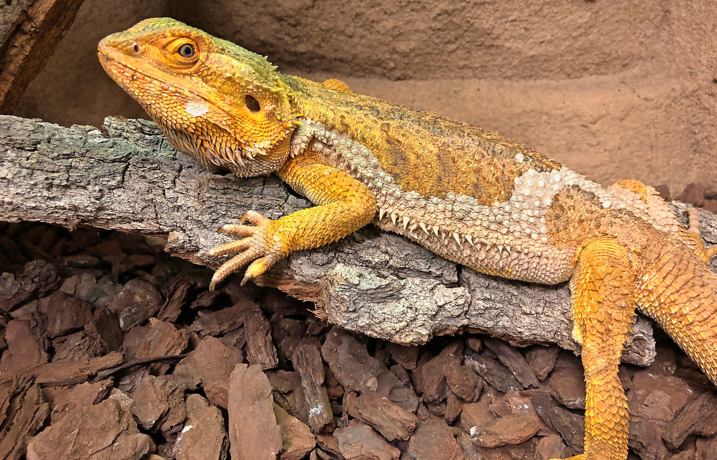
Bartagame
Reptil
Summary
Pigment-forming tumors in bearded dragonsChromatophoromas are rare tumors in reptilia. Describedin literature is one case in a bearded dragon. The present studyshows, however, that these tumors occur with higher frequencythan expected. The study aimed at characterisation of macroscopicaland clinical findings and also diagnostic methods as cytology,histopathology and immunohistochemistry. 100 growths of skinand the oral cavity of bearded dragons were investigated. 53 nonneoplasticand 47 neoplastic lesions were found. Chromatophoromas(melanophoromas n=18, iridophoromas n=4) occured with afrequency of 46% of all neoplasias. Melanophoromas were mostlygrey or black, iridophoromas were white coloured. Most chromatophoromasoccured solitarily in the skin of the body. Cytologicallyand histologically most chromatophoromas were composed ofspindle cell type neoplastic cells with varying pigmentation andsingle mitotic figures. In HE stained slides iridophores containedfine golden brown/olive green pigment granules showing typicalbirefringence when examined under polarized light. In six beardeddragons a special “mucinous type” of melanophoroma with agelatinous consistence of the cut surface characterized by looselyarranged spindle shaped cells with mild cellular and nuclear pleomorphismand large amounts of mucinous interstitial matrix wasdescribed. Signs of malignancy were: high pleomorphism, elevatedmitotic index, intravascular tumor cells and metastases. In allchromatophoroma Melan A and S100 protein were coexpressed invarying intensity and proved to be useful for diagnosing reptilianamelanotic melanophoromas. The chromatophoromas often showedmalignant behaviour. In seven cases the bearded dragons were euthanisedbecause of inoperable growth and poor prognosis. In twocases recurrence of the neoplasms was observed. According to theliterature complete surgical extirpation is the therapy of choice.
chromatophoroma
melanophoroma
iridophoroma
bearded dragon
reptile