Hemangioma in a horse
Der Praktische Tierarzt 99, 898-904
DOI: 10.2376/0032-681X-18-18
© Schlütersche Verlagsges. 2018
Publiziert: 08/2018
Zusammenfassung
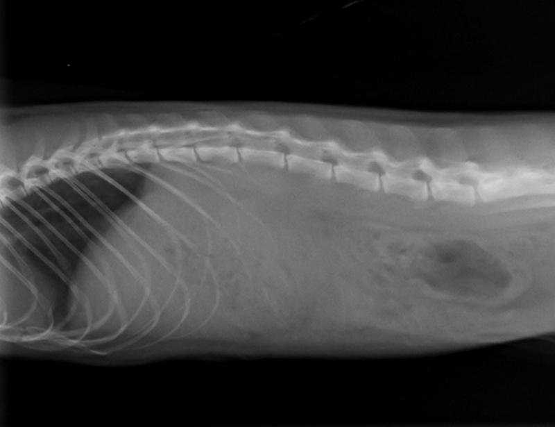
Anhand eines Fallberichts wird ein Hämangiom beim Pferd beschrieben. Hämangiome sind benigne, angiogene Neoplasien und entstehen aus entarteten Endothelzellen der Blutgefäße. Sie kommen vor allem bei Hund, Katze und Pferd vor und gelegentlich beim Schwein und den Boviden. Die kutan gelegenen Hämangiome, werden eher bei jungen Pferden an den Extremitäten beobachtet und haben eine gute Prognose bei vollständiger Entfernung. Der Jährling in dieser Studie wurde 2015 in der Klinik vorgestellt. Das Hämangiom war an der Hintergliedmaße lokalisiert und die Besitzer beschrieben ein über Wochen langsames Wachstum. Bei der klinischen Untersuchung wurde eine nicht schmerzhafte, lokal begrenzte Umfangsvermehrung palpiert, die in der Unterhaut verschieblich war. In der röntgenologischen Untersuchung konnten röntgendichte Areale dargestellt werden.
Mittels Probepunktion konnte das Hämangiom differenzialdiagnostisch von Abszessen und Seromen abgegrenzt werden, denn das Punktat zeigte frisches koaguliertes Blut. Entscheidend für den Therapieerfolg bei Vorliegen eines Hämangioms ist eine komplette chirurgische Entfernung. Diese erfolgte in Allgemeinanästhesie. Die Wundnaht verheilte komplikationslos. Die anschließende histopathologische Untersuchung erbrachte die eindeutige Diagnose eines Hämangioms. Da sich ein Hämangiom invasiv im umliegenden Gewebe ausbreiten kann, ist die Gefahr eines Rezidivs gegeben. Der Jährling wurde in den folgenden zwölf Monaten alle drei Monate kontrolliert. Dabei zeigte sich kein Rezidiv.
Summary
This case report describes the occurrence of a haemangioma in a young horse. Haemangiomas are benign angiogenic neoplasms, which arise from degenerate blood vessel endothelial cells. Haemangioma are found especially in young dogs, cats and horses; though they sometimes also occur in cattle and pigs. Subcutaneous haemangiomas tend to be more commonly found on the extremities in young horses and have a good prognosis if they can be removed completely. The yearling in this case report was presented in the clinic in 2015. The haemangioma was located on its hind leg and the horse’s owner described the tumour’s slow growth over a number of weeks. The clinical examination revealed the presence of an indolent, well-defined mass which was easily moved in the subcutis. The mass was shown as a radiopaque structure when examined radiographically. Puncture of the mass was done to differentiate it from an abscess or seroma. The fluid obtained from the mass was freshly coagulated blood. As stated previously, essential for the successful treatment of a haemangioma is its complete surgical excision. This was done under general anaesthesia.
The subsequent histopathology of the excised mass revealed it to be a haemangioma. As haemangiomas can be locally invasive, there is the danger of recurrence. Accordingly, the yearling was subsequently controlled every three months for the following 12 months. No signs of recurrence could be found. In conclusion, the diagnosis of haemangioma should always be taken into consideration with the presence of an expansive tumour and histopathology should be done to differentiate the mass from other types of tumours (e.g. haemangioepitheliomas or malignant tumours).