Kleintierpraxis 53, 141-153
© M. & H. Schaper GmbH. 2008
Publiziert: 03/2008
Zusammenfassung
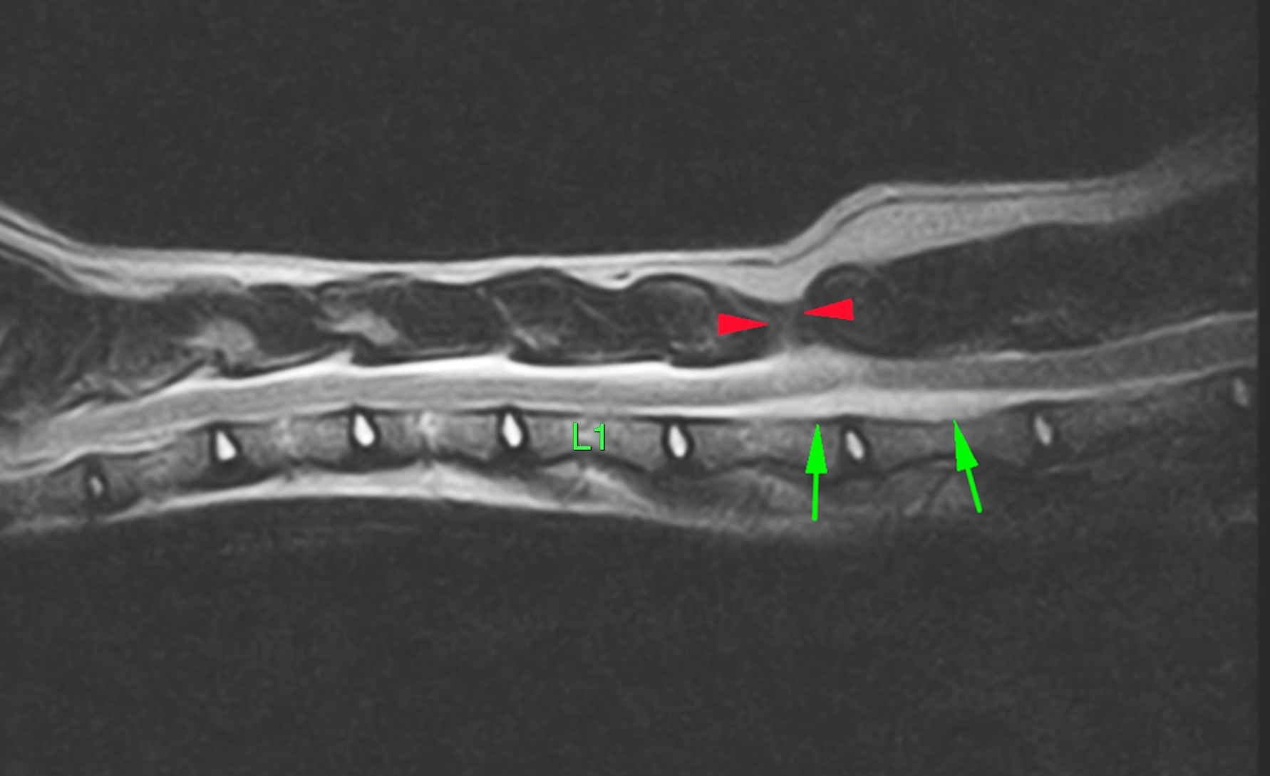
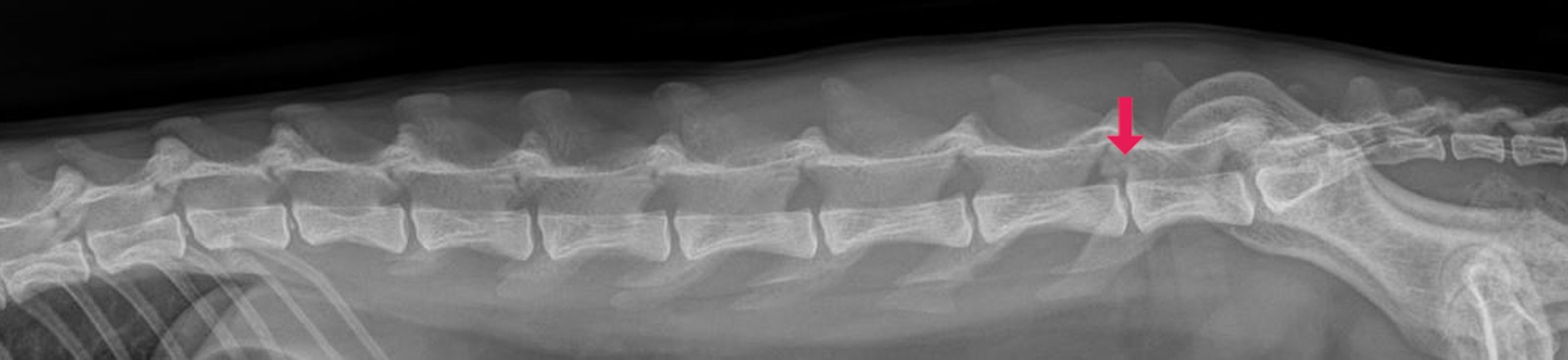
Das Ziel dieser Studie ist, eine minimalinvasive transilialeVertebralverblockung (MTV) vom 7. Lendenwirbel (L7) alsAlternative oder Ergänzung zu den bisher bestehendenOperationsmethoden zur chirurgischen Therapie von lumbosakralenStenosen darzustellen. Insgesamt wurden 33Hunde mit einem Cauda equina Kompressionssyndrom(CEKS) nach neurologischer Untersuchung, dynamischemRöntgen (Stressaufnahmen) und Magnetresonanztomographie(MRT) in zwei Gruppen unterteilt: In die Gruppe Agelangten 5 Patienten mit einer hochgradigen Diskusprotusionoder Diskusextrusion, ausgeprägter Neuroforamenstenoseund Spondylolisthesis mit Stufenbildung vom 7.Lendenwirbel (L7) zum 1. Sakralwirbel (S1). Diese wurdenmit einer partiellen Laminektomie von L7 und S1 chirurgischversorgt. Zusätzlich wurde der 7. Lendenwirbel untervisueller Kontrolle mit einem SteinmannNagelin Hyperflexiontransilial fixiert. In der Gruppe B wurde diese Technikbei 28 Hunden mit einer Spondylolisthesis, entsprechendenneurologischen Defiziten und leichter bis mittelgradigerDiskusprotusion ohne Laminektomie minimalinvasiv unterCBogenKontrolleangewandt. Nach Versorgung derPatienten in den beschriebenen Methoden konnte in derRöntgenkontrolle ein Ausgleich der Stufenbildung von L7und S1 festgestellt werden. Die vorher nach ventral disloziertenProcessi articulares craniales von S1 waren wiedernach dorsal reponiert, was eine Öffnung und Dekompressionder Neuroforamina bewirkte. In beiden Gruppen konnteeine deutliche Besserung über 95 % oder Beseitigung derklinischen Beschwerden post operationem erzielt werden.Spondylolisthesis
CaudaequinaKompressionssyndrom(CEKS)
degenerative Diskopathie
Neuroforamenstenose
Magnetresonanztomographie (MRT)
Summary
Minimal invasive transilial vertebral blockage of the 7th lumbarvertebra for the treatment of lumbosacral stenosis in the dog.The aim of this study is to present minimal invasive transilialvertebral blockage of the 7th lumbar vertebra as an alternativeor addition to the existing methods of surgical therapyof lumbosacral stenosis. A total of 33 dogs with caudaequina compression syndrome diagnosed following neurologicalexamination, dynamic Xray(stress imaging) andmagnetic resonance imaging were allocated to two groups.The five patients in Group A had a severe disc protrusion ordisc extrusion, extreme neuroforamen stenosis and spondylolisthesiswith step formation from L7 to S1. These weretreated surgically with a partial laminectomy of L7 and S1.In addition, the 7th lumbar vertebra was fixed transiliallywith a Steinmann pin in hyperflexion under visual control.In Group B, this technique was applied minimal invasivelywith Carmcontrol without laminectomy on 28 dogs withspondylolisthesis, corresponding neurological deficits andslight to moderate disc protrusion. Following treatmentof the patients with the methods described, correctionof the step formation of L7 and S1 could be ascertainedusing xraymonitoring. The cranial articular processes of S1previously dislocated ventrally were repositioned dorsallyleading to the opening and decompression of the neuroforamina.A considerable improvement of over 95% or a totalelimination of the clinical complaints was achieved postoperatively in both groups.
spondylolisthesis
cauda equina compression syndrome(CECS)
degenerative discopathy
neuroforamen stenosis
magnetic resonance imaging (MRI)