

Zusammensetzung von ovinen und caprinen Urolithen sowie kurz- und langfristiger Behandlungserfolg nach obstruktiver Urolithiasis – eine retrospektive Analyse von Patientendaten aus einer süddeutschen Tierklinik
Berliner und Münchener Tierärztliche Wochenschrift 136, 1–12
DOI: 10.2376/1439-0299-2022-25
© Schlütersche Fachmedien GmbH. 2023
Eingereicht: 20. Dezember 2022
Akzeptiert: 13. Februar 2023
Publiziert: 03/2023
Summary
Obstructive urolithiasis is common in male small ruminants. Struvite is often assumed the most frequent urolith type, but urolith analyses are rarely published, especially in Europe. Prognostic factors for short- and long-term outcomes following treatment are also rarely studied. Ninety urolith samples from small ruminants presented to a veterinary hospital were examined by infrared spectroscopy, and factors potentially associated with urolith type were statistically analysed. Treatment outcomes for 148 sheep and goats with obstructive urolithiasis were additionally assessed with particular focus on potential predictors for successful tube cystostomy (n=89) and long-term outcomes. Calcium carbonate was the most frequent urolith type, followed by silica and calcium phosphate. Species, age, animal purpose, husbandry, castration status, body condition and nutrition were significantly associated with urolith type. Goats, older animals, pets, access to pasture, castration and obesity were significantly associated with the development of calcium-based uroliths, while sheep, young age, year-round housing, uncastrated animals, non-obese body condition and high concentrate diet were associated with the development of phosphatic calculi. Of the 89 animals receiving tube cystostomy, 36 (40.4%) could be discharged from the hospital. Positive predictors for a successful short-term outcome were timely presentation for treatment, successful orthograde urethral flushing and uncompromised general condition following surgery. The recurrence rate following initially successful treatment was however high (41.3%), and statistical analyses did not identify any significant predictors for a successful long-term outcome. In accordance with previous studies, our data indicate that obstructive urolithiasis remains a disease with a guarded prognosis.
Zusammenfassung
Urolithiasis ist eine häufige Erkrankung männlicher Schafe und Ziegen. Struvit wird oft als häufigste Harnsteinart vermutet. Umfangreiche Analysen, vor allem im europäischen Raum, fehlen jedoch. Prognostische Faktoren zum kurz- und langfristigen Überleben nach der Behandlung sind nur in einzelnen Studien untersucht. Neunzig Harnsteine von kleinen Wiederkäuern, die als Patienten in eine Überweisungsklinik eingeliefert wurden, wurden mittels Infrarotspektroskopie untersucht. Faktoren mit einem möglichen Einfluss auf die Harnsteinart sowie die Überlebenschancen von 148 Schafen und Ziegen mit obstruktiver Urolithiasis wurden statistisch analysiert. Hier wurde ein besonderer Fokus auf potenzielle Einflussfaktoren für eine erfolgreiche Zystostomie mit Foley-Katheter (n = 89) und auf das langfristige Überleben gelegt.
Calciumcarbonat war die häufigste Harnsteinart, gefolgt von Silikat und Calciumphosphat. Tierart, Alter, Nutzung, Haltung, Kastrationsstatus, Übergewicht und Fütterung waren signifikant mit der Harnsteinart assoziiert. Ziegen, Hobbytiere, ältere Tiere, Weidehaltung, kastrierte Tiere und Übergewicht waren signifikant mit der Bildung von kalziumbasierten Harnsteinen assoziiert, während Schafe, jüngere Tiere, ganzjährige Stallhaltung, unkastrierte Tiere, normalgewichtige Tiere und kraftfutterreiche Fütterung mit der Bildung von phosphathaltigen Harnsteinen assoziiert waren. Nach temporärer Zystostomie mit Foley-Katheter konnten 36 von 89 operierten Tieren
(40,4 %) aus der Klinik entlassen werden. Zeitnahe Einlieferung in die Klinik, erfolgreiche orthograde Spülung der Urethra während der Operation sowie ein unbeeinträchtigtes Allgemeinbefinden nach der Operation waren positive Einflussfaktoren auf das kurzfristige Überleben. Die Rezidivrate nach erfolgreicher initialer Therapie war jedoch hoch (41,3 %) und es konnten keine signifikanten Einflussfaktoren auf das langfristige Überleben identifiziert werden. Übereinstimmend mit den Ergebnissen früherer Studien zeigen die vorliegenden Daten, dass die Prognose bei obstruktiver Urolithiasis vorsichtig ist.
Introduction
Obstructive urolithiasis is a common and frequently fatal disease in small ruminants (Hay 1990, Scully 2021). Due to the anatomic properties of the male genital tract, male animals are particularly prone to urinary obstruction, most frequently at the ischiadic arc, the sigmoid flexure or the urethral process (Sargison and Angus 2007). It is assumed that castrated males, especially those castrated at a young age, are at higher risk of developing an obstruction in comparison to intact animals due to their smaller urethral diameter (Bani Ismail et al. 2007, Radostits et al. 2007, AlLugami et al. 2017).
Multiple factors lead to supersaturation of the urine (Hay 1990, Radostits et al. 2007, Ganter 2008). Crystallization and precipitation of uroliths then follow (Defarges et al. 2020). In this process, nutritional components play an important role (Sickinger and Windhorst 2022). Pelleted, grain-based rations and a lack of roughage increase the risk of developing struvite and apatite calculi (Corbera et al. 2007, Jones and Miesner 2009). Rations high in magnesium and phosphorous also increase the risk of struvite formation (Sato and Omori 1977, Wang et al. 2009). Legume-rich rations may predispose to calcium carbonate uroliths (Jones and Miesner 2009, Smith and Sherman 2009), while plants containing oxalate promote the formation of calcium oxalate calculi (Rankins and Pugh 2012). Silica stones are frequently formed in arid regions with silica-rich soil (Smith and Sherman 2009). Reduced water intake and alkaline urine are viewed as additional risk factors for obstructive urolithiasis (Hay 1990, Radostits et al. 2007). In the United States, calcium carbonate and apatite have been reported as the most common urolith types in small ruminants in previous studies (Van Metre et al. 1996, Ewoldt et al. 2006, Osborne et al. 2009). There is, however, only very limited published information on urolith composition in small ruminants in Europe, with struvite reported as the most commonly diagnosed substance in sheep following analysis of a limited number of uroliths from Germany (Wenkel et al. 1998).
Encouraging increased water intake is a prophylactic measure applicable to all types of urinary calculi. However, different specific dietary changes are additionally necessary for the prevention of the individual urolith types. For example, in case of struvite, urinary acidification is recommended. This can be achieved by adding ammonium chloride to the diet (Stratton-Phelps and House 2004). However, permanent supplementation of this substance can lead to metabolic acidosis, followed by increased calcium release from the bones and increased renal calcium excretion. This can then create a higher risk of developing calcium carbonate uroliths (Stratton-Phelps and House 2004). Ammonium chloride supplementation would thus be counter-productive in animals with primary calcium carbonate urolithiasis. This example emphasizes the paramount importance of urolith analyses for the establishment of effective prophylactic measures, and an efficient reduction of recurrences.
Top Job:
Despite a variety of treatment approaches, obstructive urolithiasis remains a common disease with uncertain outcome. A frequently used and promising surgical treatment option is tube cystostomy with temporary implantation of a Foley catheter. Following this surgical approach, reported success rates for short-term survival ranged from 52 to 85% (Rakestraw et al. 1995, Ewoldt et al. 2006, Riedi et al. 2018b, Kretsch and Chigerwe 2021). Castration, inability to urinate despite treatment, uroperitoneum, azotaemia, hyperkalaemia and hypochloraemia have been reported to be associated with non-survival (Ewoldt et al. 2006, Riedi et al. 2018b). The analysis of long-term outcomes has often been limited by low patient numbers (Rakestraw et al. 1995, Ewoldt et al. 2006, Kretsch and Chigerwe 2021). Riedi et al. (2018b) reported that 40 of 82 animals initially treated successfully by tube cystostomy (48.8%) were still alive after 6 months, and 37 animals (45.1%) survived until one year post surgery.
This study was conducted to establish the nature of urinary calculi in sheep and goats with obstructive urolithiasis presented to a veterinary hospital in Southern Germany and to assess potential factors associated with the occurrence of various urolith types. In addition, it also aimed to generate evidence regarding prognostic factors for short- and long-term survival.
Materials and Methods
Animals and data collection
Medical records of 148 male small ruminants admitted to the Clinic for Ruminants with Ambulatory and Herd Health Services, Ludwig-Maximilians-Universität München, Germany between 2008 and 2021 with a confirmed diagnosis of obstructive urolithiasis were analysed for this study. The diagnosis was defined by the presence of the following clinical signs: inability or difficulty to urinate, straining for urine, and dilated urinary bladder or evidence of uroperitoneum. A thorough clinical examination was performed and recorded by the veterinary surgeon on duty upon arrival and a full history was taken at the time. Transabdominal ultrasonography was performed using a 5 MHz sector probe (various ultrasonic devices, e.g. HS 101V, Honda Electronics, Toyohashi, Japan). Blood samples were taken from the jugular vein into EDTA, lithium heparin, fluoride and serum tubes (Sarstedt, Nümbrecht, Germany). Information regarding species, age, breed, castration status, purpose, husbandry, nutrition, medical history, clinical signs, body condition, treatment, clinical course, blood tests, urolith analyses and short-term outcomes were gathered from the clinical records. To determine long-term outcomes, telephone interviews were performed with the owners once between two months and eight years after hospital discharge to gather information regarding the survival and potential recurrences of their animals.
Husbandry conditions were assigned to two categories: access to pasture (at least temporary/seasonal) or fully housed all year. Nutritional information was classified as either roughage-only diet, or moderate or large amounts of grain-based concentrated feed. The animals were assigned to three age categories: under 1 year, 1 to 5 years and over 5 years. Using information from the clinical records, the general condition upon arrival at the hospital was classified as follows: uncompromised, slightly compromised (tense abdominal wall or unphysiological posture, but alert), moderately compromised (reduced appetite, sounds of discomfort, straining to urinate or teeth grinding), and severely compromised (belly kicking or severe depression, recumbency). Obesity (yes/no) was a subjective assessment of the veterinarian in charge at the time of arrival. If a body condition score was recorded, animals with a BCS of four and five on a scale of one to five were classified as obese.
Laboratory analyses
Blood analyses were performed in-house using automated devices for blood gas analysis (RapidPoint 500, Siemens, Erlangen, Germany), serum biochemistry (Cobas C311, Roche, Basel, Switzerland) and haematology (HM-5, Abaxis/Zoetis, New Jersey, United States). The results were compared to published reference ranges for small ruminants (Tschuor et al. 2008).
Uroliths were submitted to a specialised commercial laboratory and examined by infrared spectroscopy (Harnsteinanalysezentrum Bonn, Germany). For statistical analyses, the different uroliths were assigned to four groups following the classification suggested by Byers (2015): calcium-based calculi (calcium carbonate, calcium oxalate), phosphatic calculi (struvite, calcium phosphate, calcium magnesium phosphate, magnesium phosphate), silica calculi and mixed/others (e.g. mixed calculi containing <80% of one component, inflammatory nidus, other material). Mixed calculi consisting of >80% of one material were assigned to the relevant groups according to their major component.
Treatment
Treatment choices followed a cascade and were always chosen in close communication with the owners. Unless an animal’s condition warranted immediate euthanasia, the first step was always an attempt to exteriorize the penis for examination following deep sedation with xylazine (sheep: 0.2 mg/kg body weight (BW) i.m.; goats: 0.05 – 0.1 mg/kg BW i.m.) and ketamine (both species: 2 – 4 mg/kg BW i.v.). If still present, the urethral process was amputated. If urinary flow was restored by this procedure, the animal remained hospitalized for between two and fourteen days for observation and medical treatment with butylscopolamine (0.4 mg/kg), non-steroidal anti-inflammatory drugs (NSAIDs) such as Meloxicam (0.5 mg/kg) or Flunixin-Meglumine (1 mg/kg) and fluid therapy, once urinary flow was restored. Antibiotics or other additional treatments were used on a case by case basis as deemed necessary by the attending veterinary surgeon. If no or insufficient urinary output was observed following amputation of the urethral process, tube cystostomy was performed immediately with the owner’s consent, or a decision for euthanasia was taken.
Surgery was performed under general anaesthesia (inhalation with isoflurane following induction with xylazine and ketamine as described above). Prior to surgery the animals routinely received antibiotics, NSAIDs and butylscopolamine, plus tetanus serum in unvaccinated cases. Tube cystostomy was performed as previously described (Rakestraw et al. 1995). Postoperative treatment included antibiotics and anti-inflammatory/analgesic drugs. Intravenous fluid therapy was administered when drainage of the urinary bladder was secured and continued on a case by case basis as long as necessary. Six to nine days following surgery the catheter was blocked for short periods to provoke urination, with the duration of the blockage gradually increased over the course of several days. The catheter was removed when the animal was able to urinate without straining, and the patient was usually discharged ten to twenty days post surgery.
Statistical analyses
Statistical analyses were performed using R version 3.6.3 (R Core Team 2021). The following factors were assessed regarding a potential association with the different urolith categories: species (sheep/goat), age, castration status (yes/no), purpose of the animal (breeding/fattening/pet), husbandry (fully housed/access to pasture), nutrition (roughage only/moderate amounts/large amounts of concentrates) and obesity (yes/no). The association between urolith categories and categorical variables were explored with Two-Sample-Chi-Square tests. The proportions within each urolith category were compared using One-Sample-Chi-Squared Goodness of Fit tests. Age was checked for normality by Shapiro-Wilk normality test. Due to not normally distributed data, urolith types in relation to age were compared by Kruskal-Wallis test, and Dunn tests were used for pairwise comparisons. P-values for multiple comparisons were corrected using the Benjamini & Hochberg method (Benjamini and Hochberg 1995).
Potential predictors for short-term survival were studied by logistic regressions. These included species (sheep/goat), castration status (yes/no), age category (<1, 1 – 5, >5 years), purpose of the animal (breeding/fattening/pet), duration of disease prior to presentation (<1, 1 – 2, >2 days), blood values on admission (creatinine, urea, potassium, chloride, sodium), successful urethral flushing during the operation (yes/no) and general condition following surgery (uncompromised/compromised/severely compromised). Only animals which received tube cystostomy and thus best possible treatment were included in these analyses to exclude any bias by owners deciding to avoid surgery for reasons of cost.
Analysis of long-term survival was performed for all animals discharged from the hospital using a Cox proportional hazards model (Cox 1972) and included the following potential predictors: species (sheep/goat), castration status (yes/no), age, obesity (yes/no), purpose of the animal (breeding/fattening/pet), husbandry (fully housed/access to pasture), complications on arrival (yes/no), type of treatment (no treatment/conservative/surgery) and successful urethral flushing during the operation (yes/no).
The statistical analyses were limited to univariate models. Multivariate models were attempted but failed due to limited case numbers within some categories.
Results were considered significant for p ≤ 0.05; p>0.05 and ≤0.1 was considered a tendency.
Results
Patient characteristics and descriptive results
Within the study period, 148 animals (79 goats and 69 sheep) fulfilled the inclusion criteria. The majority of these animals (102 animals; 68.9%) were between one and five years old, and 94 animals (63.5%) were kept as pets. Access to pasture was the predominant husbandry system (125 animals; 84.5%). Detailed descriptive results for all 148 animals are shown in table 1.
The clinical findings at the initial examination are summarized in table 2. Only 44.6% (66/148) of the animals showed obvious signs of pain and distress at hospital admission. Urination was impossible for 75% (111/148). The most frequently recorded abnormality during the initial examination was a dilated urinary bladder, as seen in 89.8% (114/127) of the animals with available ultrasonography results.
Before arrival at the hospital, 63.5% (94/148) of the patients had been treated by a referring veterinarian. Administered treatments by the referring veterinary surgeons included NSAIDs (47 animals), antibiotics (25 animals), spasmolytic drugs (51 animals), amputation of the urethral process (14 animals) and homoeopathy (6 animals). Twenty-nine animals had been misdiagnosed as cases of indigestion or other gastrointestinal problems and had been treated accordingly.
Laboratory analyses
Blood analyses upon arrival at the hospital were performed for 139 of the 148 animals (93.9%), but not all individual blood values were available for all patients due to presentation during emergency hours. Elevated creatinine levels were present in 97.0% (131/135) of the studied animals, with measurements ranging from 48 mmol/l to 2670 mmol/l (mean: 660 mmol/l; median: 471 mmol/l; reference range: 39–104 mmol/l; Tschuor et al. 2008). Urea levels were elevated in 85.2% (115/135) of the patients and ranged from 2.6 mmol/l to 113.5 mmol/l. The mean (29.9 mmol/l) and the median (22.8 mmol/l) urea levels were also distinctly higher than the reference ranges (2.1–7.2 mmol/l; Tschuor et al. 2008). Decreased serum chloride levels were also a common feature and present in 67.6% (92/136) of the animals with available results. Chloride values ranged from 13 mmol/l to 119 mmol/l (mean: 96 mmol/l; median: 98 mmol/l; reference range: 102–117 mmol/l; Tschuor et al. 2008). Serum calcium levels were decreased in 99.3% (136/137) of the patients, with the mean (1.04 mmol/l) and median (1.07 mmol/l) values well outside the reference range (2.2–2.8 mmol/l; Tschuor et al. 2008). In addition, mean and median blood sodium levels (both 141 mmol/l) were slightly lower than the reference range (147–159 mmol/l; Tschuor et al. 2008), while these values were above the reference range for creatine kinase (CK) (mean: 836 mmol/l; median: 362 mmol/l; reference range: 86–268 mmol/l; Tschuor et al. 2008). No distinct deviations of the mean and median from the reference ranges were noticed for the remaining blood values (phosphorus, potassium, PCV, pH, bicarbonate).
Urolith analyses
Uroliths were analysed from 90 animals (49 goats, 41 sheep). The most common urolith type was pure calcium carbonate, followed by pure silica and pure calcium phosphate. Detailed results are shown in table 3, and assignment to the various urolith categories (Byers 2015) is presented in table 4. Six cases of pure calcium phosphate, four cases of amorphous calcium phosphate and one case of carbonated apatite are summarized under calcium phosphate.
The eight cases of mixed calcium carbonate included four cases of 80% calcium carbonate plus 20% amorphous calcium phosphate, three animals with 90% calcium carbonate plus 10% amorphous calcium phosphate, and one case of 60% calcium carbonate plus 40% silica. The three cases of mixed silica included one animal with 60% silica plus 40% calcium oxalate, one with 90% silica plus 10 % calcium oxalate and another with 80% silica plus 20% calcium carbonate. The three cases of mixed struvite included one case of 60% struvite plus 40% silica, one of 80% struvite plus 20% protein and a third of 60% struvite plus 40% calcium phosphate. There was one case of 90% calcium oxalate mixed with 10% silica.
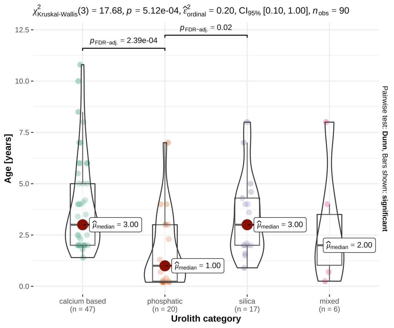
The urolith composition differed between species (p = 0.002). Calcium-based (p = 0.03) and silica calculi (p = 0.09) were more frequent in goats, while phosphatic concrements were more frequent in ovine patients (p = 0.007). The age of the animals was also significantly associated with the urolith type (p < 0.001). Animals with calcium based uroliths (p < 0.001) and silica uroliths (p = 0.02) were significantly older than animals with phosphatic calculi. Details are shown in figure 1.
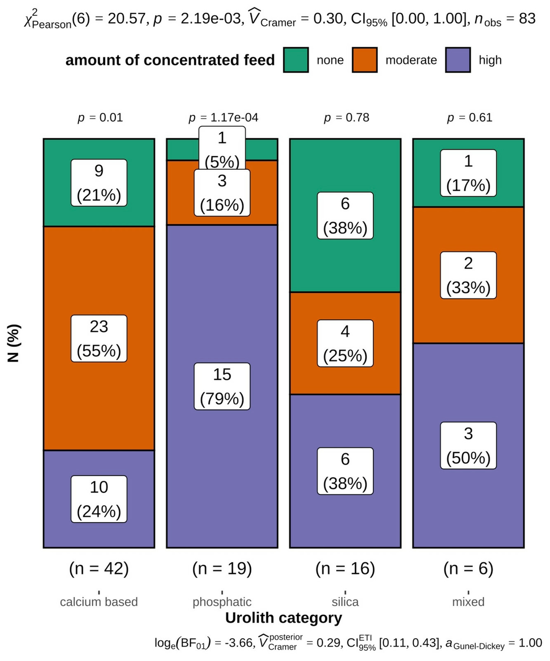
Further factors significantly associated with urolith category were castration status (p < 0.001), obesity (p = 0.003), purpose (p < 0.001), husbandry (p < 0.001) and nutrition (p = 0.002). A significantly higher proportion of castrated animals had calcium-based (p = 0.002) or silica calculi (p = 0.008), while more animals with phosphatic calculi were intact (p < 0.001). A higher proportion of obese animals were diagnosed with calcium-based uroliths (p = 0.001), while more animals with normal body condition presented with phosphatic calculi (p = 0.04). Pets were over-represented in the calcium-based (p < 0.001) and silica (p < 0.001) urolith categories. All nine animals kept for fattening were diagnosed with phosphatic concrements. Pasture-based husbandry was more frequently observed for animals with calcium-based (p < 0.001) and silica uroliths (p < 0.001). Animals with phosphatic calculi more frequently received high concentrate rations (p < 0.001) (Fig. 2).
Treatment and short-term outcomes
Twenty-nine of the 148 submitted animals (29/148; 19.6%) were euthanized immediately following confirmation of the diagnosis without any treatment, based on their owners’ decisions or in case of particularly poor prognosis. Five animals (5/148; 3.4%) received medical treatment only. Twenty-five animals (25/148; 16.9%) were treated by amputation of the urethral process without further surgery. A total of 89 animals (89/148; 60.1%) received tube cystostomy. This included 49 animals treated by amputation of the urethral process followed by tube cystostomy. In 20 animals it was not possible to extract the penis for amputation of the urethral process either due to adhesions between penis and prepuce following very early castration, or due to severe urinary oedema or inflammation. These animals thus received tube cystostomy only. For another 20 animals no information was available from the clinical records regarding extraction of the penis or amputation of the urethral process prior to surgery.
During tube cystostomy, orthograde flushing of the urethra was attempted in 72 of the 89 cases (72/89; 80.9%). Flushing was not attempted in some very young or very small animals due to a lack of a suitably sized catheter. Urethral flushing was successful in 27 animals (27/72; 37.5%; 24 completely, 3 partially) and not possible in 45 cases (45/72; 62.5%).
Free fluid was discovered in the abdominal cavity during surgery in 44 of the 89 surgical cases (44/89; 49.4%), and adhesions due to intraabdominal inflammatory processes were discovered in 14 animals (14/89; 15.7%).
Five animals died during anaesthesia and thirteen were euthanized during surgery due to the presence of severe complications. Seventy-one of the 89 operated animals (71/89; 79.8%) initially survived surgery. Following the operation, 26 of these 71 animals (26/71; 36.6%) showed good general demeanor, 28 (28/71; 39.4%) had reduced general demeanor and 16 (16/71; 22.5%) showed obvious signs of distress and pain. One animal died shortly after the operation, so its post-operative general condition could not be fully assessed. Including this case, a total of five animals which initially survived surgery (5/71, 7.0%) died before the first blocking attempt of the catheter, i.e. prior to day 7 post surgery, and another seven (7/71; 9.9%) were euthanized during this period. Of the 59 animals (59/71; 83.1%) surviving until the first blocking attempt, thirty-six (36/59; 61.0%) were able to urinate at the first attempt. Three of these were however later euthanized due to other complications such as severe cystitis and/or peritonitis. The remaining 23 animals (23/59; 39.0%) were unable to urinate at the first blocking attempt. Three of these later successfully passed urine at follow-up attempts and survived, while the remaining 20 were eventually euthanized as a result of continued urethral obstruction. In total, thirty-six animals (36/89; 40.4%) with tube cystostomy were discharged.
Across all treatment groups, 91 of the 148 animals (65.5%) were euthanized at the hospital, eleven (7.4%) died during their stay and 46 animals (31.1%) were discharged alive. Details of short-term survival within the various treatment groups and reasons for euthanasia are listed in tables 5, 6 and 7.
Potential predictors for short term survival following tube cystostomy
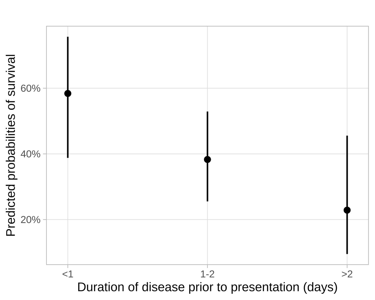
Owners’ decisions against surgery were a major factor influencing short-term outcomes. Potential predictors for short-term survival were therefore only examined for the 89 animals receiving tube cystostomy and thus full treatment. Species, age, castration status, purpose, blood sodium and blood potassium levels were not significant in the univariate logistic regressions and were therefore dismissed as potential predictors for a successful outcome. However, univariate logistic models identified the duration of disease prior to presentation (p = 0.03, Fig. 3), high blood urea levels (p = 0.004; Fig. 4a), high creatinine (p = 0.014; Fig. 4b) and decreased blood chloride (p = 0.004; Fig. 4c) as significant predictors for decreased survival, and successful urethral flushing (p < 0.001) and uncompromised general condition following surgery (p = 0.001) as influential predictors for a successful outcome. Animals that had been ill for more than two days prior to presentation were less likely to survive compared to animals that were presented for treatment the day the first symptoms occurred (OR: 0.21; p = 0.017). Animals with successful urethral flushing had higher chances of survival compared to animals with unsuccessful flushing attempts during surgery (OR: 5.88; p < 0.001; CI: 2.27–21.6). Animals which showed uncompromised general condition following surgery were more likely to survive compared to those with a compromised (OR: 0.18; p = 0.004; CI: 0.05–0.59) or severely compromised (OR: 0.04; p < 0.001; CI: 0.01–0.2) general condition. Animals with higher blood urea levels (p = 0.01) and higher blood creatinine levels (p = 0.028) were less likely to survive. Lower chloride levels were associated with a lower chance of survival (p = 0.009).

Long-term outcome
Of the 46 animals (30 goats, 16 sheep) discharged alive from the hospital, 19 had recurrent obstructions (19/46; 41.3%). Sixteen of these had one recurrence between seven days and 18 months after hospital discharge, while three animals had two recurrent obstructions after 9 and 45 months, after 12 and 42 months and after 9 and 21 months, respectively. The median time between hospital discharge and first recurrence was 9 months. Fifteen animals were euthanized due to the first recurrence, while one goat was successfully operated a second time, but lost to further long-term follow-up after this successful second tube cystostomy. Of the three animals suffering two known recurrences, all three were successfully operated a second time for their first recurrence. Two were then euthanized due to their second recurrence. One sheep was operated three times and survived both recurrences. This animal was eventually euthanized 2.5 years after the third operation due to arthritis. Five animals were euthanized without a recurrence due to other diseases. Two animals died following hospital discharge. No long-term outcome could be determined for five animals. Fifteen animals survived without a recurrence during an observation time between 6 months and 3.5 years, but we were unable to identify similarities in these fifteen cases. Univariate Cox proportional hazards models did not identify any significant predictors for a successful long-term outcome.
Discussion
This retrospective cohort study included small ruminant patients from the German regions of Bavaria and Baden-Württemberg as well as occasional patients from neighbouring Austria. Due to this regional limitation, and a pre-selection of animals submitted to a veterinary hospital for confirmation of diagnosis and treatment, the results cannot be considered representative for the entire German sheep and goat population. While it is well known that obstructive urolithiasis is a common disease in grain-fed, fattening lambs in preparation for slaughter (Jones and Miesner 2009, Rankins and Pugh 2012), animals kept for this purpose were under-represented in our study cohort. Owners of pets or valuable breeding animals are more likely to present their animals for potentially lengthy and expensive treatments, and particularly pets were over-represented, an observation which is in accordance with previous studies carried out in hospital settings (Osborne et al. 2009, Gamsjäger and Chigerwe 2021). Despite these limitations, this study includes the highest number of urolith analyses from small ruminants in Germany to date, and indeed in Europe, with the vast majority of previous studies originating from the USA or other overseas countries (Manning and Blaney 1986, Osborne et al. 2009, Jones et al. 2017, Gamsjäger and Chigerwe 2021).
The most frequent urolith type in our study cohort was calcium carbonate in both small ruminant species. Studies from the USA showed similar results (Osborne et al. 2009, Gamsjäger and Chigerwe 2021). These authors explained the predominance of calcium carbonate uroliths with an over-representation of pets in their study population, and this was also the case in our studied animals. It is therefore highly likely that other urolith types are more frequent in the German small ruminant population than their observed proportions in the studied patients. This assumption is supported by one of the few previous German studies on this subject, which – in contrast to our results – reported struvite in 24 of 31 examined sheep (77.4%), and calcium phosphate uroliths in the other seven (22.6%) (Wenkel et al. 1998). The authors did not report any details regarding nutrition, age or purpose of these animals, so it can only be assumed that the difference in predominant urolith type between this study and our results may be due to differences in patient pre-selection, and a potential over-representation of animals on high concentrate rations in the sheep examined by Wenkel et al. (1998).
Urolith analyses were not carried out in a comparable case load treated by two Swiss veterinary hospitals (Riedi et al. 2018a). However, 34 of 56 radiographs (60.7%) taken in this study showed radiopaque calculi. The most frequent urolith types in this study cohort are therefore most likely radiopaque substances such as calcium carbonate, calcium oxalate or silica (Videla and van Amstel 2016, Riedi et al. 2018a).
One study from Texas, USA, reported amorphous magnesium calcium phosphate (AMCP) as the most frequent urolith type (Jones et al. 2017). Most animals in their study population were used for exhibition purposes (19 of 49 animals) or breeding (7 of 49 animals), while only 18 animals were kept as pets. These had significantly higher odds of developing calcium carbonate uroliths compared to exhibition animals. The differences in study population, and potentially in nutritional and geographic circumstances, may explain why AMCP was not commonly observed in our study cohort.
In our patients, phosphatic calculi were significantly more frequent in sheep, young and uncastrated animals, patients with normal body condition, and animals receiving high concentrate rations. All these parameters describe the population of sheep kept for fattening purposes, or young breeding rams raised on high concentrate rations in preparation for sale. These rations are usually high in grain-based components and low in roughage, thus high in phosphorous and low in calcium, predisposing to the formation of struvite and other phosphatic calculi (Hay 1990, Smith and Sherman 2009). Statistical analyses could only be performed independently for each factor due to relatively low case numbers and the high number of potentially influential factors, so interactions or the relative importance of each individual parameter could not be statistically assessed. It can however be assumed that nutrition most likely plays a more important role than other factors such as castration status, husbandry, body condition or species, since 15 of 18 animals with phosphatic uroliths were fed a high concentrate diet. As 33 of 47 animals (70.2%) receiving a high amount of concentrated feed were sheep, and 34 of the 41 sheep (82.9%) were intact, the parameters species, castration status and nutrition are not truly independent. The same limitation applies to factors associated with other urolith types: calcium based uroliths were more frequently identified in goats, older animals, castrated animals, pets, animals with access to pasture and overweight animals. Many of these parameters describe the typical population of goats kept as pets. The relative importance of each individual parameter could not be assessed. Age has however also been identified as a potentially influential factor for an increased risk of calcium-based urolithiasis by a number of other authors in a variety of species such as goats, sheep, dogs and man (Wisener et al. 2010, Nwaokorie et al. 2015, Jones et al. 2017, Hunprasit et al. 2019, Katz et al. 2021). In contrast, a potential influence of castration on urolith type is controversial. One study reports significantly higher odds of developing calcium carbonate uroliths for castrated animals (Nwaokorie et al. 2015), while other authors found no significant association between castration status and urolith type (Jones et al. 2017). The overall risk for obstructive urolithiasis, irrespective of urolith type, is however likely to be higher for castrated males, particularly for animals castrated prior to sexual maturity, because of a smaller urethral diameter and remaining preputial adhesions (Belonje 1965, Bani Ismail et al. 2007, Sickinger et al. 2019).
Nutrition is also likely to play an important role for calcium based uroliths, and consumption of large amounts of calcium containing legumes has been suggested as an aetiological factor for the formation of calcium carbonate calculi by Jones and Miesner (2009). However, pasture conditions and roughage sources in Southern Germany typically consist of unimproved meadows and permanent grassland, which may naturally contain a certain amount of clover, but other legumes such as alfalfa are not commonly used as a major feed component. It is therefore unclear whether the natural clover content of unimproved grassland is sufficient to cause calcium carbonate urolithiasis, or if there are additional, potentially unknown factors involved. The potential role of obesity, purpose (pets) and species in the development of calcium carbonate uroliths still needs to be fully established, but several authors have observed a high frequency of calcium carbonate calculi in goats. Jones et al. (2017) and Van Metre et al. (1996) found calcium carbonate uroliths only in goats. Goats of African descent have been reported to have higher odds of developing calcium carbonate uroliths but were over-represented in the respective study (Nwaokorie et al. 2015). Whether there is indeed a species effect remains to be determined.
Of the 89 animals receiving tube cystostomy, 36 could be discharged alive (40.4%). This short-term survival rate is similar to a recent Swiss study with a comparable study population, which reported a short-term success rate of 52% (Riedi et al. 2018b), and to a different German study reporting a surgical success rate of 5 out of 14 animals (35.7%) (Dühlmeier et al. 2007). It is however noticeably lower than several reports from the USA, which reported short-term survival rates of 76.2% (Ewoldt et al. 2006), 80% (Rakestraw et al. 1995) and 84.3% (Gamsjäger and Chigerwe 2021). The lower survival rates in our study can be explained by the fact that some studies either excluded animals in which establishing urethral patency was impossible at the first admission (Gamsjäger and Chigerwe 2021) – in contrast to the present study, which included all animals that received tube cystostomy irrespective of the severity of their condition – or contained lower case numbers (Rakestraw et al. 1995). Facilities in the United States may also see more primary cases with a shorter duration of disease. This has been shown to be associated with a better outcome (Riedi et al. 2018b). Ninety-four of the 148 animals in the present study (63.5%) had been treated prior to referral to the hospital, some of which for prolonged periods. The clinical signs for obstructive urolithiasis can vary widely and may be unspecific, or not always immediately obvious (Riedi et al. 2018a). Very close monitoring of the animals is therefore essential in order to identify the disease at an early stage. More subtle signs may go unnoticed, especially in larger groups, or may be masked by stressful events. For instance, 53.1% (76/143) of the studied animals showed no obvious signs of pain or distress at hospital admission, but subtle signs are very likely to have been masked by the stress of transport. The rate of misdiagnosis by initially attending veterinary surgeons was relatively high (29/148; 19.6%), with clinical signs such as reduced appetite, reduced ruminal motility and even straining commonly mistaken for signs of gastrointestinal problems. Early diagnosis is however crucial for best possible outcomes, and particularly ultrasonography is a useful tool to confirm the diagnosis. A dilated urinary bladder is a reliable indication for urinary obstruction (Scott 2000). This was also reflected in our study population, with a dilated bladder (without or with additional free abdominal fluid) present in 123 of 127 animals (96.9%) with available ultrasonography results. Blood analyses can also be useful for diagnostic purposes, as serum urea and creatinine were elevated in 85.2% (115/135) and 97.0% (131/135) of the submitted animals with available blood analyses. However, it needs to be borne in mind that blood values can be unchanged in very early stages (Riedi et al. 2018b). Very high serum urea and creatinine levels were significantly associated with non-survival. Urea and creatinine levels rise with prolonged duration of disease (Fortier et al. 2004, Riedi et al. 2018b). The duration of disease is thus most likely the true factor influencing treatment outcomes, with blood values reflecting prolonged periods of urinary obstruction. According to Dühlmeier et al. (2007), prognostic statements based on creatinine or urea levels should and cannot be made. Hypochloraemia was also significantly associated with non-survival. In conjunction with hyponatraemia it can be an indication of bladder rupture (Donecker and Bellamy 1982, George et al. 2007). Animals with ruptured bladders were often euthanized during surgery, their outcomes were therefore worse than for animals with intact bladders.
Although castration is considered a risk factor for developing obstructive urolithiasis (Radostits et al. 2007, Smith and Sherman 2009), the association of castration with treatment outcomes remains unclear. Some authors report higher survival rates for castrated animals (Ewoldt et al. 2006), while others have seen higher survival rates for intact animals (Riedi et al. 2018b). Castration status was not significantly associated with short term survival in our study cohort, a finding supported by the results of Kretsch and Chigerwe (2021).
A cautiously positive short-term prognosis can be made if orthograde urethral flushing is possible during the operation, and if the animals show good general demeanour following surgery. Unsuccessful urethral flushing however does not necessarily predict a negative outcome (Jones et al. 2012). Predicted probabilities of short-term success were however significantly reduced for these animals in our study cohort.
No predictors for a successful long-term outcome could be identified, and long-term prognosis remains very guarded due to a high recurrence rate of 41.3% (19/46). Recurrence rates in previous studies were variable and ranged from 7.7% (Rakestraw et al. 1995) to 40.2% (Gamsjäger and Chigerwe 2021). These partly lower recurrence rates may be influenced by lower patient numbers (Rakestraw et al. 1995, Iselin et al. 2001, Fortier et al. 2004, Ewoldt et al. 2006), or short observation times (Rakestraw et al. 1995, Ewoldt et al. 2006). The lower values in many previous studies are therefore likely to be an under-estimation. Our own results regarding the long-term outcomes also need to be treated with caution and are indeed likely to be an under-estimation. It was not possible to repeatedly follow up the vast majority of the animals during their entire lifetime. In addition, the time between surgery and follow-up call was highly variable between animals and as short as 2 months in some individual cases due to presentation only months prior to the end of the observation period for this study. Finally, not all owners could be reached by telephone, and their animals were thus lost to follow-up. It is therefore highly likely that some recurrences did not come to our attention, thus leading to a likely under-estimation of the recurrence rate.
In conclusion, urolith analysis is necessary in order to provide tailored advice regarding preventive nutritional management, since calcium based and silica calculi were more frequent than anticipated or suggested by many sources, and measures instigated to prevent phosphatic calculi are unlikely to be successful in these cases. Obstructive urolithiasis remains a disease with uncertain outcome, and owners and first opinion veterinarians must be made aware of the varying and often subtle clinical signs in order to ensure timely presentation for surgery, thus increasing treatment chances. During and immediately following the operation, successful orthograde urethral flushing and good general condition following surgery can be seen as positive prognostic indicators. The overall prognosis however remains guarded, particularly in the long term, as recurrences are frequently observed.
Ethical approval
The authors hereby declare that they have followed the universally accepted guidelines of good scientific and good veterinary practice while preparing the present paper. This non-invasive field study did not include any procedures requiring formal ethical approval.
Conflict of interest statement
The authors hereby declare that they have no proprietary, professional or other personal interests in any product, service and/or company that could have influenced the contents or opinions expressed in this publication.
Funding
Not applicable.
Authors contribution
Conceptualization: KV.
Methodology: KV, YZ, LE.
Data collection, analysis and interpretation: LE, KV, YZ.
Statistical analyses: YZ.
Visualization: YZ.
Writing – original draft preparation: LE..
Writing – review and editing: KV.
Critical revision: YZ, GKS.
Supervision, KV, GKS.
All authors have read and agreed to the final version of the manuscript.
Address for correspondence
Katja Voigt, Ludwig-Maximilians-Universität München, Klinik für Wiederkäuer mit Ambulanz und Bestandsbetreuung, Sonnenstr. 16, 85764 Oberschleißheim, katja.voigt@lmu.de
References
AlLugami A, von Pückler K, Wehrend A (2017): Sonography of the distal urethra in lambs. Acta Vet Scand 59(16): 16–16.
Bani Ismail Z, Al-Zghoul M, Al-Majali A, Khraim N (2007): Effects of castration on penile and urethral development in Awassi lambs. Bulg J Vet Med 10(1): 29–34.
Belonje PC (1965): Observations on the post natal development of the penis in merino ram lambs and wethers: the possible relationship to the passage of urinary calculi. J S Afr Vet Assoc 36(3): 381–383.
Benjamini Y, Hochberg Y (1995): Controlling the false discovery rate: a practical and powerful approach to multiple testing. J R Stat Soc Series B Stat Methodol 57(1): 289–300.
Byers SR (2015): Urolithiasis. In: Smith B, Van Metre DC (eds.), Large animal internal medicine. 5th ed. Elsevier, St. Louis, MO, 897–903.
Corbera JA, Morales M, Doreste F (2007): Experimental Struvite Urolithiasis in Goats. J Appl Anim Res 32(2): 191–194.
Cox DR (1972): Regression Models and Life-Tables. J R Stat Soc Series B Stat Methodol 34(2): 187–220.
Defarges A, Evason M, Dunn M, Berent A (2020): Urolithiasis in Small Animals. In: Bruyette D (ed.), Clinical Small Animal Internal Medicine. John Wiley & Sons, Hoboken, NJ, 1123–1156.
Donecker JM, Bellamy JE (1982): Blood chemical abnormalities in cattle with ruptured bladders and ruptured urethras. Can Vet J 23: 355–357.
Dühlmeier R, Zibell G, von Altrock A, Roth C, Schröder C, Thies K, Ganter M (2007): Urolithiasis beim kleinen Wiederkäuer – Behandlungsmethoden und klinische Rekonvaleszenz. Tierarztl Prax Ausg G Grosstiere Nutztiere 35(03): 175–182.
Ewoldt JM, Anderson DE, Miesner MD, Saville WJ (2006): Short- and Long-Term Outcome and Factors Predicting Survival After Surgical Tube Cystostomy for Treatment of Obstructive Urolithiasis in Small Ruminants. Vet Surg 35(5): 417–422.
Fortier LA, Gregg AJ, Erb HN (2004): Caprine Obstructive Urolithiasis: Requirement for 2nd Surgical Intervention and Mortality After Percutaneous Tube Cystostomy, Surgical Tube Cystostomy, or Urinary Bladder Marsupialization. Vet Surg 33(6): 661–667.
Gamsjäger L, Chigerwe M (2021): Risk factors for, frequency, and type of complications after temporary tube cystostomy in goats, sheep, and pigs. Vet Surg 50(2): 283–293.
Ganter M (2008): Urolithiasis. In: Behrens H, Ganter M, Hiepe T (Hrsg.), Lehrbuch der Schafkrankheiten. 4. Aufl. Parey, Berlin, 37–41.
George JW, Hird DW, George LW (2007): Serum biochemical abnormalities in goats with uroliths: 107 cases (1992–2003). J Am Vet Med Assoc 230(1): 101–106.
Hay L (1990): Prevention and treatment of urolithiasis in sheep. In Pract 12(3): 87–91.
Hunprasit V, Schreiner PJ, Bender JB (2019): Epidemiologic evaluation of calcium oxalate urolithiasis in dogs in the United States: 2010–2015. J Vet Intern Med 33(5): 2090–2095.
Iselin U, Lischer CJ, Braun U, Steiner A (2001): Zystotomie mit und ohne temporärer Implantation eines präpubikalen Ballonkatheters zur Behandlung der obstruktiven Urolithiasis beim kleinen Wiederkäuer: eine retrospektive Studie. Wien Tierarztl Monatsschr 88(2): 39–45.
Jones ML, Miesner MD (2009): Urolithiasis. In: Anderson DE, Rings DM (eds.), Food Animal Practice. Elsevier, St. Louis, MO, 322–325.
Jones ML, Miesner M, Baird AN, Pugh DG (2012): Diseases of the Urinary System. In: Pugh DG, Baird AN (eds.), Sheep and goat medicine. 2nd ed. Elsevier/Saunders, Maryland Heights, MO, 325–360.
Jones ML, Gibbons PM, Roussel AJ (2017): Mineral Composition of Uroliths Obtained from Sheep and Goats with Obstructive Urolithiasis. J Vet Intern Med 31(4): 1202–1208.
Katz JE, Soodana-Prakash N, Jain A (2021): Influence of Age and Geography on Chemical Composition of 98043 Urinary Stones from the USA. Eur Urol Open Sci 34: 19–26.
Kretsch CM, Chigerwe M (2021): Assessment of preoperative and postoperative l-lactatemia and clinical outcomes in goats undergoing tube cystostomy: A retrospective study of 34 cases (2015–2020). J Vet Intern Med 35: 2926–2932.
Manning RA, Blaney BJ (1986): Identification of uroliths by infrared spectroscopy. Aust Vet J 63(12): 393–396.
Nwaokorie EE, Osborne CA, Lulich JP (2015): Risk factors for calcium carbonate urolithiasis in goats. J Am Vet Med Assoc 247(3): 293–299.
Osborne CA, Albasan H, Lulich JP (2009): Quantitative analysis of 4468 uroliths retrieved from farm animals, exotic species, and wildlife submitted to the Minnesota Urolith Center: 1981 to 2007. Vet Clin North Am Small Anim Pract 39(1): 65–75.
R Core Team (2021): R: A language and environment for statistical computing. R Foundation for Statistical computing, Vienna, Austria.
Radostits OM, Gay CC, Hinchcliff KW, Constable PD (2007): Veterinary Medicine. Elsevier, New York.
Rakestraw PC, Fubini SL, Gilbert RO (1995): Tube cystostomy for treatment of obstructive urolithiasis in small ruminants. Vet Surg 24: 498–505.
Rankins DLJ, Pugh DG (2012): Feeding and Nutrition. In: Pugh DG, Baird AN (eds.), Sheep and goat medicine. 2nd ed. Elsevier/Saunders, Maryland Heights, MO, 18–49.
Riedi AK, Knubben-Schweizer G, Meylan M (2018a): Clinical findings and diagnostic procedures in 270 small ruminants with obstructive urolithiasis. J Vet Intern Med 32(3): 1274–1282.
Riedi AK, Nathues C, Knubben-Schweizer G (2018b): Variables of initial examination and clinical management associated with survival in small ruminants with obstructive urolithiasis. J Vet Intern Med 32(6): 2105–2114.
Sargison ND, Angus KW (2007): Diseases of the urinary system. In: Aitken ID (ed.), Diseases of sheep. 4th ed. Blackwell Publishing, Oxford, 395–402.
Sato H, Omori S (1977): Incidence of Urinary Calculi in Goats Fed a High Phosphorus Diet. Nihon Juigaku Zasshi 39(5): 531–537.
Scott P (2000): Ultrasonography of the urinary tract in male sheep with urethral obstruction. In Pract 22(6): 329–333.
Scully CM (2021): Management of Urologic Conditions in Small Ruminants. Vet Clin North Am Food Anim Pract 37(1): 93–104.
Sickinger M, Windhorst A (2022): A systematic review on urolithiasis in small ruminants according to nutrition-dependent prevalence and outcome after surgery. Vet World 15(3): 809–817.
Sickinger M, Allugami A, von Pückler K (2019): Comparative ultrasonographic examination and measurements of the urethra and penis of castrated and intact male lambs. Pol J Vet Sci 22(1): 127–132.
Smith MC, Sherman DM (2009): Goat medicine. John Wiley & Sons, Ames, IA.
Stratton-Phelps M, House L (2004): Effect of a commercial anion dietary supplement on acid-base balance, urine volume, and urinary ion excretion in male goats fed oat or grass hay diets. Am J Vet Res 65(10): 1391–1397.
Tschuor, Riond, Braun, Lutz (2008): Hämatologische und klinisch-chemische Referenzwerte für adulte Ziegen und Schafe. Schweiz Arch Tierheilkd 150(6): 287–295.
Van Metre DC, House JK, Smith B, Thurmond M, George LW, Angelos SM, Angelos JA, Fecteau G (1996): Obstructive urolithiasis in ruminants: surgical management and prevention. Compend Continu Educ Vet 18: 275–289.
Videla R, van Amstel S (2016): Urolithiasis. Vet Clin North Am Food Anim Pract 32(3): 687–700.
Wang J-Y, Sun W-D, Wang X-L (2009): Comparison of effect of high intake of magnesium with high intake of phosphorus and potassium on urolithiasis in goats fed with cottonseed meal diet. Res Vet Sci 87(1): 79–84.
Wenkel R, Berg W, Prange H (1998): Harnsteine bei Kleintieren und anderen Tierarten. Eine retrospektive Studie aus den Jahren 1980–1989. Dtsch Tierarztl Wochenschr 105(5): 182–186.
Wisener LV, Pearl DL, Houston DM (2010): Risk factors for the incidence of calcium oxalate uroliths or magnesium ammonium phosphate uroliths for dogs in Ontario, Canada, from 1998 to 2006. Am J Vet Res 71(9): 1045–1054.

Kostenfreier Download
Klicken Sie hier, wenn Sie das PDF BMTW-10.2376-1439-0299-2022-25-Eicher.pdf (0.31 MB) herunterladen möchten
Kostenfreier Download
Klicken Sie hier, wenn Sie das PDF BMTW-10.2376-1439-0299-2022-25-Eicher-Tabelle1.pdf (0.11 MB) herunterladen möchten
Kostenfreier Download
Klicken Sie hier, wenn Sie das PDF BMTW-10.2376-1439-0299-2022-25-Eicher-Tabelle2.pdf (0.1 MB) herunterladen möchten
Kostenfreier Download
Klicken Sie hier, wenn Sie das PDF BMTW-10.2376-1439-0299-2022-25-Eicher-Tabelle3.pdf (0.1 MB) herunterladen möchten
Kostenfreier Download
Klicken Sie hier, wenn Sie das PDF BMTW-10.2376-1439-0299-2022-25-Eicher-Tabelle4.pdf (0.11 MB) herunterladen möchten
Kostenfreier Download
Klicken Sie hier, wenn Sie das PDF BMTW-10.2376-1439-0299-2022-25-Eicher-Tabelle5.pdf (0.1 MB) herunterladen möchten
Kostenfreier Download
Klicken Sie hier, wenn Sie das PDF BMTW-10.2376-1439-0299-2022-25-Eicher-Tabelle6.pdf (0.1 MB) herunterladen möchten
Kostenfreier Download
Klicken Sie hier, wenn Sie das PDF BMTW-10.2376-1439-0299-2022-25-Eicher-Tabelle7.pdf (0.11 MB) herunterladen möchten


